“A little duloxetine to loosen my tongue”

The drug duloxetine, marketed under the brand name Cymbalta, was originally developed for depression or anxiety but is now prescribed mostly for pain management.  An article from neurologists in Russia and Malaysia now reports that three people with PSP who were receiving duloxetine for their mood issues experienced important, unexpected improvement in their speech. 

The article’s first author is Dr. Oybek Turgunkhujaev of the Semeynaya Clinic in Moscow and its senior author is Dr. Shen-Yang Lin of Universiti Malaya in Kuala Lumpur.  Unfortunately, the journal, Movement Disorders Clinical Practice, is not open-access and because it’s only a brief article, there is no abstract in PubMed for me to link to.

The mechanism of action duloxetine is to enhance the action of the neurotransmitters serotonin and norepinephrine by blocking the brain cell’s ability to re-absorb those molecules from the synapse.  That’s a standard way for brain neurons to regulate and sharpen its signals to other neurons.  That process is called “reuptake” and you’ve probably heard of the SSRIs or selective serotonin reuptake inhibitors. Duloxetine works on norepinephrine as well as serotonin, so it’s an SNRI.   Other SNRIs in common use include venlafaxine (Effexor) and desvenlafaxine (Pristiq). The figure below shows the mechanism of action of duloxetine and venlafaxine. 

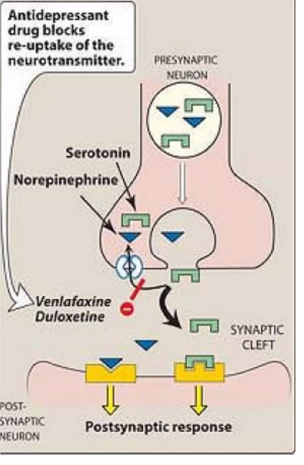
Nerd alert: This mostly takes place in the pons, specifically the locus ceruleus (for norepinephrine), raphe nuclei (for serotonin), but to a lesser extent in prefrontal cortex of the cerebrum (both norepinephrine and serotonin).

The journal article is accompanied by before-and-after videos of two of the three patients.  They all had difficulties not only with slurring, but also with difficulties initiating speech and stuttering.  The delay to onset of the drug’s benefit is described only as “within weeks.”  All three patients also experienced an improvement in their mood, which could have been a direct effect of the drug on the natural reaction to their improved ability to speak.

The authors do a good job of describing how the drug’s known mechanism of action might address the parts of the brain that when impaired, are known to cause those symptoms.  But they also recognize that the general alerting and energizing effect of duloxetine could explain the result as well.  Placebo effect seems unlikely, as the durations of benefit were too long: “> 3 months,” “15 months until death” and “>5 months.” 

Bottom line: This is encouraging and I hope further reports of duloxetine in PSP from other centers are forthcoming. BUT . . . and MEANWHILE . . .

Physicians considering prescribing duloxetine off-label for speech problems in PSP should be aware that it can have important side effects and drug/dietary interactions.  I can’t get into those here, but they are described in detail in the 31-page FDA-approved package insert and its one-page summary. Needless to say, there has been no formal trial of duloxetine in PSP, so that population could have different/additional/worse side effects relative to the mostly younger people in the depression and pain trials on which the package insert was based. 

Posts from an outpost

A colleague and co-author of mine, Michiko Kimura Bruno, MD, has started a blog that might interest you. Its platform is the Psychology Today site and its very cool overall topic is neurological disorders from the psychological standpoint. Dr. Bruno alerted me to this because the third post is about PSP.

Dr. Bruno is an academic movement disorders specialist, like me, but in Hawaii, not New Jersey. (No jokes, please.) She works at The Queen’s Medical Center, which is affiliated with the John A. Burns School of Medicine at The University of Hawai’i at Mānoa, where she is Professor of Neurology. She directs CurePSP’s Center of Care at her institution. To date, Dr. Bruno is author or co-author of eight peer-reviewed papers on PSP, five of which are about its social or psychological aspects. Take a look.

Michiko K. Bruno, MD

PSP meets metaphenomic annotation

Back in the 19th Century, diagnosing brain disorders relied mostly on listening carefully to the patient’s history, performing a detailed neurological examination, and knowing what previous, similar patients turned out to have at autopsy.  That approach remains important, but since the early 20th Century, chemical, electrical and imaging tests have replaced it to a great extent.  But now, neurologists at one of the world’s most prestigious medical centers, the Queen Square Institute of Neurology at University College London, have turned back the clock.

The researchers were motivated by the failure to date of modern techniques short of autopsy to provide a sufficient level of diagnostic certainty for the atypical Parkinsonian disorders.  So, they squeezed some more value out of the traditional approach.  First, they reviewed 125 publications since 1992 describing a total of 5,748 people with Parkinsonian disorders of various types and a diagnostic answer at autopsy.  Those proven diagnoses were Parkinson’s disease (30%), PSP (23.5%) multiple system atrophy (16.8%), dementia with Lewy bodies (6.0%), corticobasal degeneration (4.6%).  There was also a large group called “others” (19.6%), who mostly had Alzheimer’s disease, which in its advanced stages can acquire stiffness and slowness and look “Parkinsonian.”  Keep in mind that the sources of these numbers were specialized academic referral centers, where the atypical Parkinsonian disorders would be over-represented.

Led by first author Dr. Quin Massey and senior author Dr. Christian Lambert, the researchers invented a new statistical technique called “metaphenomic annotation,” which involved feeding the 125 journal articles into commercially available text-mining software to tabulate the patient’s lists of symptoms against the autopsy results.

Etymology lesson of the day: “Meta” means “beyond,” in this case beyond the confines of a single published study.  “Pheno” means “outward appearance,” in this case the features of the disease during life.  “Phenomics” means a large collection of such features, analogous to “genomics” as a large collection of genes. “Annotation” in this case means somehow managing to shoehorn all that data from disparate sources into a usable database.

They found that among the 1,195 people with PSP diagnosed during life, the autopsy diagnosis was PSP in 89.5%, PD in 3.0%, CBD in 2.5%, MSA in 1.3%, and “other” in 3.7%. Among the 1,349 people whose autopsy showed PSP, the diagnosis during life was PSP in 79.3%, PD in 8.5%, MSA in 5.6%, CBS in 3.6%, DLB in 0.2% and “other” in 2.7%.  The graph below shows the corresponding numbers for all five conditions and the “others” group. The curving bands show how the diagnoses made during life (the “clinical cohort” on the left) were corrected by autopsy to the diagnoses (the “histological cohort” on the right).  (“Histology” is the study of body tissues through the microscope.)

Figure and table from: Quin Massey, Leonidas Nihoyannopoulos, Peter Zeidman, Thomas Warne, Kailash Bhatia, Sonia Gandhi, Christian Lambert. Refining the diagnostic accuracy of Parkinsonian disorders using metaphenomic annotation of the clinicopathological literature. NPJ Parkinsons Disease 2025 Nov 10;11:314.

If you think the figure above is information-rich but a bit confusing, the table below may be more of both.  Each of the five rows is labeled with a disease defined by autopsy, and each column represents how that disease compares with each of the other four with regard to diagnostic signs during life.  For example, for assistance in distinguishing PSP from MSA, look at the third row, second column, where square waves (an abnormal eye movement) are listed as being more common in PSP.  The number in parentheses is the ”positive likelihood ratio” for distinguishing PSP from MSA.  The PLR is calculated as (sensitivity / (1-specificity)) and can be informally described for this example as how much more likely is PSP than MSA to be the correct diagnosis, based on just this one diagnostic feature.

This analysis can be very useful for the vast majority of neurologists and mid-level neurological professionals unfamiliar with the details of the differences among the atypical Parkinsonian disorders.  Even experienced movement disorders neurologists will find it useful to have all this information in one place and neatly quantified.

A great part of this project is that the researchers have made the annotated data and their computer code publicly available for the use of other researchers.

Jesse Jackson

The Rev. Jesse L. Jackson, Sr., the civil rights leader and political activist, has announced that his 2017 diagnosis of Parkinson’s disease has been changed to PSP.

I have no inside information on the details of Rev. Jackson’s condition, but I can tell you that this is a common scenario for people with the second-most-common type of PSP, called PSP-Parkinsonism (PSP-P).  It accounts for about 15% to 35% of all PSP, depending on the source of the data, and starts out looking like Parkinson’s disease (PD), typically with asymmetric stiffness of the limbs and often a tremor, without much cognitive, eye movement or balance difficulty.   The symptoms, like those of PD, usually respond to levodopa, though not quite as well or for as many years. 

PSP-P progresses at a rate typical for PD, not the faster rate for most types of PSP.  The average lifespan of PSP-Richardson syndrome (PSP-RS), which accounts for about 45% to 55% of PSP, is about 6 years, while for PSP-P, it’s 9 years and for PSP overall, 7 years.  (The lifespan of people with PD receiving modern treatment averages about 16 years, but many neurologists today don’t know that before levodopa became available in 1970, it was about 9 years, like PSP-P.)

The typical journey of someone with PSP-P starts with a satisfactory response to levodopa.  But the dosage requirement increases more rapidly and balance problems develop sooner than in PD.  After a few years, the neurologist may suspect one of the “atypical Parkinsonian disorders,” examine the patient more closely than at the previous follow-up visits, find the eye movement problem and frontal cognitive issues of PSP, and change the diagnosis.  Often, this re-evaluation is performed by a second neurologist whose opinion was sought by the patient or family member worried about the atypical symptom course and unsatisfactory levodopa response.

So, if Rev. Jackson does have PSP-P, he would now be at about the average (i.e., the median) survival duration.  Of course, about half of all people with PSP-P survive past the median, some for as long as a total of 20 years or even more.

A scientific comment: Keep in mind that PSP-P wasn’t described in the medical literature until 2005 and formal criteria to distinguish among the various (now 10) different PSP subtypes didn’t appear until 2017.  Before then, most people with PSP-P would not have fulfilled the existing formal criteria for PSP, which were published back in 1996.  Therefore, pre-2017 statistics on the prevalence or lifespan of people with PSP refer mostly to PSP-RS, which is the name given in 2005 to what was defined as “PSP” by the 1996 criteria.

Another scientific comment: PSP-RS, PSP-P and the other 8 subtypes all have the same set of microscopical changes in the brain, featuring neurofibrillary tangles of tau protein and tufted astrocytes.  It’s just the sets of locations around the brain that differ to some extent.  We don’t yet know what causes this “cell-type specificity,” but the best theory is that it’s slight differences in the mis-folding pattern of tau, caused by slightly different locations of abnormal attachment of phosphate groups on the protein.

And finally, an editorial comment: Almost all clinical drug trials in PSP only accept participants with PSP-RS.  There are several good reasons for that, but that policy excludes half of all those with PSP, raising the theoretical possibility that any resulting treatment may work only in that sub-type.  I’ll discuss potential solutions in a future post.

Meanwhile, my deepest gratitude to Rev. Jackson for sacrificing some degree of personal medical privacy in the service of increasing public awareness of PSP and my best wishes to him in the journey forward.

Suggestive eyes

The current diagnostic criteria for PSP were published in 2017. Unlike the previous standard set of PSP criteria from 1986, this one defined not only “possible PSP” and “probable PSP,” but also a new, lower level of certainty called “suggestive of PSP.”  That applied to people with PSP features insufficient to satisfy the “possible” criteria.  It was designed to maximize sensitivity at the likely cost of specificity.  Both the 1986 and 2017 sets of criteria defined “definite PSP” as requiring autopsy confirmation.

For PSP-Richardson syndrome, the most common of the 10 PSP subtypes, the critical difference between “suggestive of” and “possible” relates to eye movements. The former has no restriction or slowing of eye movement, but does have either square-wave jerks (rapid, irregular, low-amplitude, horizontal jerks of the eyes when staring straight ahead, separated by very brief pauses) or difficulty opening the eyes (also called “blepharospasm”).  Both “suggestive of” and “possible” PSP-Richardson also require at least a bit of balance loss.

The PSP research group in Barcelona, one of the world’s leading such projects, maintains a longitudinal registry and observational study.  Like most longitudinal studies, it gathers detailed information about the participants at the time of enrollment and periodically thereafter, seeking ways to use one to predict the other — diagnostic and prognostic markers. 

The group’s latest project was headed by Celia Painous, MD PhD, a post-doc working under senior author Yaroslau Compta, MD PhD.  It enrolled 131 participants with PSP, 12 with Parkinson’s disease and 18 healthy people as controls.  Of those with PSP, 23 had “suggestive of” criteria for one of the PSP subtypes (mostly PSP-Parkinsonism) and 108 met “possible” or “probable” criteria (mostly PSP-Richardson syndrome).

The testing included not only the standard history and physical exam for PSP but also a blood test for neurofilament light chain (NfL) and several tests in spinal fluid:  alpha-synuclein seed amplification assay (asyn-SAA), neurofilament light chain (NfL), total tau (t-tau), tau with a phosphate at amino acid 181 (p-tau), and beta amyloid (Aβ42).  All of these have been proposed as diagnostic markers for PSP or other tauopathies.

The testing also included something unusual for this type of project – a precise test of eye movement termed “video-oculography.” The participant sits comfortably at a desk, resting chin and forehead against rigid supports, as at the optometrist’s office.  They perform  visual tasks shown on a computer screen while a video camera records the movements. The images below are from a technical article on the device.  If you read it, keep in mind that it was written by the manufacturer’s employees as a marketing tool.

The visual tasks used a bright dot in the middle of the screen as a starting point and other dim dots that could light up.  For most of the tests, the person was instructed to look as quickly as possible from the starting point to other dots as they lit up in turn.  The device measured delays in starting the movement, speed, undershoot, and overshoot.  The procedure also required the participant to perform “anti-saccades,” where they’re told to move their eyes not to the dot that just lit up, but to a different dot that remains dim, situated in exactly the opposite direction from the starting point.  This can be performed in any of the four directions of gaze. It tests the ability to follow instructions and to resist one’s natural inclinations, both of which are impaired early in PSP and other disorders of the frontal lobes.

Sure enough, compared with the subjects with PD and the controls, the group with “suggestive of” PSP at the first visit showed more errors than the PD or healthy control groups in the anti-saccade task in the vertical directions, with greater delay in responding and slower movement.  It’s true that the duration of the “suggestive” PSP symptom(s) in that group was four years.  Still, the fact that only very minimal signs of PSP were present shows that the disease was in a very mild stage.  We can therefore hope that a harmless, painless, inexpensive, sensitive test for abnormalities on the anti-saccade task could provide a very early marker for PSP.  

Before long, we may see such a test added to research protocols seeking people with PSP before clear, overt symptoms develop.  That would be very useful in studies of genetic or environmental risk factors, where groups with and without the risk factor are compared with regard to the incidence of PSP.  It’s likely that for work with PSP, video-oculography could be combined with other markers to provide fewer false positives.