Bad for the goose, good for the gander?

A disturbing piece of news this week about an influential 2006 paper in Nature about Alzheimer’s disease.  Turns out it was likely that some of the data in the published version were deliberately faked.  The paper was about beta amyloid, which is not an issue in PSP.  In fact, this could actually be good news for PSP research.  Here’s why:

In the experiments reported in the 2006 paper, researchers at the University of Minnesota Twin Cities used mice carrying a copy of the human amyloid precursor protein (APP) gene with a mutational variant known to cause AD in humans.  (In the normal human brain, the protein product of the APP gene is cut to form beta-amyloid, abbreviated, “A-beta.”)  The researchers allowed the mice to develop cognitive deficits, analyzed their brains, and found a type of small A-beta aggregates never before seen, dubbing them “A-beta*56.”  They extracted the small aggregates, called “oligomers,” and injected them into the brains of genetically normal (“wild-type”) rats, which proceeded to develop AD-like cognitive disabilities. 

Ever since A-beta was identified as a critical player in AD in 1984, researchers had been trying to nail down just what form that protein takes in the process of causing, or contributing to, the disease.  The 2006 paper seemed finally to answer that question and formed the basis for innumerable subsequent experiments world-wide and hundreds of millions of dollars spent by the NIH, philanthropies and drug companies to build upon it in pursuit of an AD treatment. 

The 2006 Nature paper used a commonplace lab technique called Western blot to separate out different proteins from a mixture.  A bit of the mixture is placed on a flat layer of absorptive material and subjected to an electrical field.  The heavier proteins move more slowly than the lighter ones.  The resulting array is exposed to an antibody-based stain that allows it to be seen.  The positions and sizes of the individual protein spots are then analyzed. 

A sample Western blot unrelated to the research discussed here. The numbers on the left are the molecular weights of the proteins in kiloDaltons (one Dalton is the weight of one proton). The leftmost column shows the separation of a standard mixture of proteins of known weight as a benchmark. The other two columns show components of a protein mixture before (middle) and after (right) the addition of an enzyme that cuts proteins.

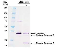
But now, a whistleblower has reported evidence that some of the Western blot images in the publication and many others from the same lab were placed where they didn’t belong, citing faint lines between blots that could result from cutting-and-pasting.  There was also an instance of two blots with identical size and shape, something with a likelihood approaching zero absent a copy-and-paste operation.  The journal Science hired two scientists unconnected to the Minnesota team to take a look.  They confirmed that deliberate falsification is highly likely, though there’s no smoking gun, which would require access to the original Western blot images or to the original data readouts. Nor, so far, has there been a confession. 

Meanwhile, what’s the upshot?  For the AD field, it means that the treatment trials of anti-A-beta drugs were based on much less laboratory evidence than was thought, possibly explaining why they all failed.  (Aducanumab, the antibody approved in 2021 by the FDA, targets A-beta, but its clinical benefit is highly controversial, Medicare refuses to cover the treatment, and most neurologists opt not to prescribe it.)  That means that by default, anti-AD treatments addressing tau, the other protein aggregating in AD, deserve more attention.

Some experts have questioned the importance of A-beta in AD for decades, but only in the last 15 years or so has AD research into tau as the alternative received serious support.  In PSP, tau is the only protein that consistently aggregates and there’s no evidence of A-beta misbehavior at all.  PSP is therefore considered by many scientists to be a good test bed for anti-tau treatments for AD.  That’s why I think that if these new doubts about A-beta in AD direct attention to tau, an intensification of tau-based PSP research could result, and that could, by extension, benefit AD as well. 

While two anti-tau antibodies have failed to slow the progression of PSP in clinical trials, there are many other ways to address tau in PSP, including one trial currently recruiting and at least two more set to start in the next year. 

So, let’s hope that this week’s revelation gives PSP research a boost and AD research a long-overdue redirection.


Here’s a detailed editorial in Science explaining all of this (without my own speculation about the possible benefit for PSP research). But it’s behind the journal’s paywall and I didn’t want to post the pdf I have access to through my university. That would be another form of dishonesty.

Dr. John Steele, fondly remembered

The PSP community mourns the passing of neurologist John C. Steele, MD on May 21, 2022, surrounded by his children in Bali, Indonesia, his most recent home. He was 87.

Dr. Steele was one of the physicians at the University of Toronto who in 1963 and 1964 published the defining clinical and pathologic descriptions of PSP.  The eponym, “Steele-Richardson-Olszewski syndrome” predominated in the medical literature for decades and is still used by some writers to honor the accomplishment. At the time, Dr. Steele was a neurology resident, Dr. J.C. Richardson was his mentor and department chief, and Dr. Jerzy Olszewski was a neuropathologist. Their 1964 paper in Archives of Neurology remains today the most frequently cited article on PSP not just for its primacy, but also for the thoroughness of its clinical and pathologic details.

Son and grandson of physicians, Dr. Steele was born in Toronto in 1934. He earned undergraduate and medical degrees at the University of Toronto and completed a neurology residency at Toronto General Hospital in 1965. He married Margaret Porter, an artist and writer who authored a children’s book on PSP.  Dr. Steele is survived by children Alex, Erica and Julia and grandchildren Jonathan, Sophia and Sean.

After his training, Dr. Steele won the prestigious McLachlan Fellowship, allowing him to pursue studies in Britain and France for two years. From that point on, his career was unconventional. He spent a year practicing and teaching in Thailand, returning in 1968 to the University of Toronto as a pediatric neurologist. In 1972, he moved to the Pacific, where he would spend the rest of his life.

He first worked as a general physician on the remote atoll of Majuro in the Marshall Islands doing everything from delivering babies to removing fishhooks, sailing on small freighters to deliver medical care on even more remote atolls.  After six years in Majuro, Dr. Steele spent a year at the London School of Hygiene and Tropical Medicine for fellowship training in clinical tropical medicine. He then moved to the island of Pohnpei in the Eastern Caroline Islands, where he trained local doctors and nurses under a clinical appointment on the faculty at the University of Hawaii Medical School.  From Pohnpei he also continued his work of practicing medicine in remote islands.

In 1982 he settled on the island of Guam as the neurologist at the US Navy’s base hospital and medical director of the local VA clinic. On Guam and nearby islands Dr. Steele cared for and studied individuals with a neurodegenerative disorder endemic to the indigenous Chamorro people. It was called lytico-bodig or the ALS-parkinsonism-dementia complex (PDC). Over the years, Dr. Steele invited and hosted multiple scientists to study this geographically and ethnically specific disorder. He spoke at numerous international medical and epidemiological conferences to create interest in PDC among researchers. Perhaps his most famous scientific guest on Guam was Dr. Oliver Sacks, whose 1997 book “The Island of the Colorblind” features a detailed portrait of Dr. Steele and his work. (The title refers not to Guam, but to the atoll of Pingelap, 1,000 miles east, where a different disease is endemic.)

Despite the lack of formal research facilities on Guam, Dr. Steele found ways to collaborate with other scientists in state-of-the-art inquiries into the cause of PDC. His warm relationship with the community as a local physician provided access to information on traditional practices, helping to elucidate risk factors for the development of the disease. He assisted in constructing detailed family trees to couple with modern molecular genetics performed by collaborators.  Those relied on Dr. Steele’s having accomplished the difficult and delicate task of securing consent for blood samples and brain autopsies. Those studies ultimately showed that mutations in genes previously known as risk factors for other neurodegenerative diseases are over-represented in the PDC population but do not fully explain its cause. This raises the possibility of as-yet-unsuspected genes or of toxic or infectious contributors.

Dr. Steele’s insights into the Chamorro’s dietary habits helped form the hypothesis that PDC was caused by a toxin in the fruit of a cycad tree, the “false sago palm,” by consuming fruit bats (“flying foxes”) that eat the fruit and bioconcentrate its toxins. One such toxin, beta-Methylamino-L-alanine (BMAA), is produced by cyanobacteria in the trees’ roots. The toxic mechanism of this amino acid remains unclear but may rely on its mis-incorporation into proteins in place of serine, thereby encouraging misfolding of the resulting protein. Another compound in the same fruit, beta-D-glucoside, acts as an excitotoxin at glutamate receptors, another mechanism known to cause brain degeneration.

Favoring the fruit bat hypothesis is the observation that PDC has slowly disappeared over the decades since World War II, as traditional dietary practices gave way to Westernization of the Chamorros’ lifestyles. Dr. Steele’s indefatigable work with the Chamorro population was instrumental in this idea, which today remains one of the leading non-genetic hypotheses explaining PDC.

A major inspiration for Dr. Steele in his work on Guam was the similarity between PDC and PSP. In 1963, during his neurology residency in Toronto, his department hosted visiting lecturer Dr. Asao Hirano, a leading neuropathologist who had studied PDC in the 1950s. At that visit, Dr. Hirano examined the brain specimens from the original PSP patients and was struck by the similarity with PDC. Twenty years later, soon after arriving on Guam, Dr. Steele saw a similarity of the impaired downward eye movement and other outward features in the two diseases. Although PSP occurs world-wide and differs from PDC in important molecular details, Dr. Steele recognized that their similarities could prove key. He approached the puzzle of PDC by continually probing the rapidly accumulating knowledge of PSP and by collating the theories and data of a wide array of specialists. As he pointed out many times, the comparative study of PSP and PDC may shed light not only on those two disorders, but also on all neurodegenerative diseases. He framed his life’s work and scientific aspirations in that way.

Few of us can claim to have set so worthy a goal or to have accomplished as much in its service.

Simple but effective

A chance re-encounter

A 2019 article I came across this week dragged me back into blog posting after a month-long break (sorry, fans — I have no excuse).  I remember seeing the paper at the time but blew it off as mere confirmation of previous publications.  But it actually may provide a way to diagnose PSP years before symptoms appear. 

The problem is a familiar one

As you know from my constant harping on the subject, what we really need are two things: a way to diagnose PSP in its earliest stages, preferably before it causes any disabling symptoms (or any symptoms at all); and a way to prevent the disease process from progressing further than that.  In official lingo: a marker and neuroprotection. 

All sorts of marker proposals are showing promise: leading the pack right now are tests of blood or spinal fluid for neurofilament light chain or tau, PET scans for tau, and various MRI techniques.  Two of the more distant contenders are smartphone-based eye movement measurements and skin biopsies for tau aggregates. The problem is to differentiate very early PSP from normal aging and from competing diagnostic possibilities such as Parkinson’s, MSA and dementia with Lewy bodies. 

Get out your rulers

MRI measurements of the volume of the cerebrum is a very sensitive way to track the progression of PSP and is used in drug trials routinely to compare the rate of brain loss in the treatment group to that in the placebo group.  But it doesn’t work for diagnosing the disease in the first place.  For that, you need to image a part of the brain that, unlike the cerebrum, is involved early in the course of the disease.  It also has to be easy to image using standard MRI machines.  The dorsal midbrain does both. 

As an internal comparator, the study also measured the size of the pons, which is the segment of brainstem just below the midbrain.  It atrophies little in PSP.  For both measures, they used the area in square centimeters of the structure on a mid-sagittal MRI cut (one that slices the head perfectly into left and right halves).  See the image below.

MRI in the mid-sagittal plane, with nose at left of each. The left image shows the dorsal midbrain and the right, the pons. a radiologist drew the outlines by hand with a mouse. The MRI machine’s software calculates the area with each outline in square centimeters. (From Cui et al BMC Neurology 2020)

Now, while the dorsal midbrain is where vertical eye movement, the hallmark of PSP, is situated, it’s not where PSP starts.  That happens in subthalamic nucleus, the globus pallidus and the substantia nigra.  But the dorsal midbrain gets involved soon enough, is much easier to image than those things, and is consistently involved in the classic form of PSP, Richardson syndrome. 

History is not bunk

So, with that as background, Dr. Jong Hyeon Ahn and colleagues from six university hospitals in South Korea found 27 patients with PSP with brain MRIs not only after their PSP symptoms began, but also before they began.  The scans had been performed for non-PSP symptoms such as transient dizziness, fainting, suspected stroke, or headache.  In fact, the article says that elderly South Koreans often request — and receive — brain MRIs as part of their routine checkups.  (Who knew?)  The MRIs were routine, with none of the standardization across radiology sites that are commonplace in multi-center drug studies.  In other words, these were “real world” MRIs.

The pre-symptomatic MRIs were performed an average of 28 months (range: 12-48 months) before PSP symptoms began and the researchers pored over their records to make sure there were no symptoms at the time suggestive of PSP.  They rejected MRIs done within 12 months of symptom onset to further reduce the chance that the symptoms prompting the scan were part of PSP.

They compared these pre-symptomatic MRIs to the same patients’ post-onset MRIs and to those of 27 patients with Parkinson’s and another 27 with no known brain disorder.  The 27 with PSP all had the classic PSP-Richardson syndrome, where the vertical eye movement problem is more prominent than in the less common PSP subtypes. 

I few paragraphs ago, I mentioned that the pons was also measured.  In some diseases, both the midbrain and pons atrophy together, but only in PSP is the midbrain affected far worse.  So they divided the areas of the pons by that of the midbrain, expecting that ratio to be higher in PSP than in competing diagnostic possibilities. 

The results

The graph below compares the four subject groups by their pons area, midbrain area and pons/midbrain ratio. There’s some overlap between groups, but the averages (the means) differ both for the midbrain alone and for the pons/midbrain ratio.  The horizontal bars with asterisks indicate a statistically significant difference between the means of two groups at the ends of the bar.  The pons alone showed no differences, as expected, but the midbrain alone did show a difference and the pons/midbrain ratio did even better than that.

Areas of dorsal midbrain and pons as measured on mid-sagittal MRI. The horizontal brackets with asterisks indicate statistically significant differences between groups. P=pons, M=midbrain, RS=Richardson’s syndrome, PD=Parkinson’s disease (from Ahn et al Park Rel Dis 2022)

Those differences weren’t just at the level of the group means, which would be scientifically interesting but close to useless for patient care.  For the pons/midbrain ratio, the accuracy (the fraction of subjects correctly classified by the test) for pre-symptomatic PSP vs PD was 89% and for pre-symptomatic PSP vs controls, it was 93%.  A more critical statistic from the standpoint of avoiding false positives is the specificity, which for the pons/midbrain measurement comparing PSP and PD, was an amazing 100%.  It was the same for the PSP vs controls. 

Receiver operating curves showing the trade-off between the sensitivity and specificity of the midbrain area (blue) with pons/midbrain ratio (green) in distinguishing patients with PSP from those with Parkinson’s disease (left two graphs) or controls (right two graphs). The two upper graphs compare pre-symptomatic PSP with PD or controls. The two bottom scans compare post-onset PSP with PD or controls. (From Ahn et al. Park Rel Disord 2022)

Now — for the green eyeshades

A strength of the study is that all the pre-symptomatic MRIs were more than 1 year before symptoms began.  Any shorter than that would raise questions of whether very subtle PSP features might have been present.  Another strength is that the MRIs were performed on ordinary machines available in any radiology office.

One caveat is that all 27 PSP patients had the PSP-Richardson form, and the findings may not apply to PSP-Parkinsonism or the other atypical forms.  Another is that the patients were alive and not autopsy-confirmed in their diagnoses and a third is that the neurological evaluations had been performed by general neurologists rather than by movement disorder specialists.

The take-home

So, we await confirmation by other researchers with larger subject numbers and comparisons of PSP with MSA and DLB.  We also need to standardize the measurement of the pons and midbrain areas to strengthen the real-world diagnostic value of this painless, harmless and apparently highly accurate test.  Coupling this test with other simple ones may create an even more accurate diagnostic battery.

This could be a keeper.  Then all we’ll need is a way to keep everyone pre-symptomatic.