Imaging in the diagnosis of PSP

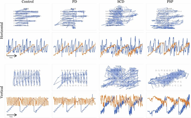
A very active area of research right now is how various imaging techniques (“scans” in English) can and cannot assist in distinguishing the atypical Parkinsonian disorders from other conditions and from one another.  Yes, this is important for clinical care and counseling.  But even more important right now is that until we have specific treatments for these diseases, we need accurate diagnosis in living people.  This is important for laboratory researchers who want to know the true diagnosis of the patient who supplied a fluid sample, and to the designers of clinical trials who want to make sure the patients in their trials have the disease for which the treatment was designed. 

Dr. Jennifer L. Whitwell, a radiology researcher at the Mayo Clinic in Rochester, MN has just published a very helpful review of that topic in Current Opinion in Neurology.  It gets pretty technical, but here are the takeaways with, of course, my own editorial contributions:

  • Measurement of atrophy by MRI: 
    • The magnetic resonance Parkinsonism index (MRPI), especially its updated version, the MRPI 2.0, gives excellent differentiation of PSP from non-PSP, with an area under the receiver operating curve of 0.98. (That statistic is 1.00 for perfect accuracy and 0.50 for no better than a flip of a coin. For a bit more explanation of the AUROC and a graph, see this post from last year.) 
    • Routine MRI can also be used to compare the various PSP variants with regard to atrophy of specific brain structures.  Atrophy of the brainstem is worse in PSP-Richardson syndrome, PSP-CBS and PSP-frontal than in the others.  This information could be useful in treatment trial design because some PSP variants progress faster than others. So, a trial of a disease-slowing treatment could potentially determine which patients have which variants and adjust the statistical analysis of the treatment outcomes accordingly.
    • MRI shows that atrophy in PSP-Richardson’s syndrome starts in the midbrain, (the upper part of the brainstem where the substantia nigra and the vertical gaze centers reside), followed by a succession of other areas of the brainstem, cerebellum and basal ganglia, before finally reaching the cerebral cortex.  But two PSP subtypes involving relatively more cortical function (the frontal behavioral type and the corticobasal syndrome type) spread into the frontal cortex much earlier in the process, although like PSP-RS, they start in the midbrain.  These observations could help guide designers of other imaging-based diagnostic tests for PSP.
  • Measurement of metabolism by fluorodeoxyglucose positron emission tomography (FDG PET):  The pattern of reduced brain tissue metabolic activity in PSP can be distinguished from normal with an AUROC of 0.99.  Distinguishing PSP from corticobasal syndrome, multiple system atrophy and Parkinson’s disease is more difficult but still useful at 0.90.
  • Measurement of tau deposition by PET:  A PET technique that reveals the location of abnormal tau in Alzheimer’s is already in standard clinical use for AD, but it doesn’t work well for other tauopathies.  Two techniques designed for PSP are expected to enter pivotal clinical trials in the next few months.  In small studies, the two ligands called PI-2620 and APN-1607 (formerly called PM-PBB3) have shown good results in distinguishing PSP from the other tau disorders and Parkinsonian disorders.  (A PET ligand is the chemical injected intravenously that sticks to the brain chemical of interest, allowing its location to be mapped.) But these two ligands can occasionally give conflicting results when given to the same patient, so the AUROCs from the upcoming trials are eagerly awaited.
  • Measurement of iron deposition by MRI: This technique, called quantitative susceptibility mapping (QSM), is not part of a routine MRI, but it can be performed with the same machine.  Its PSP-diagnostic results are not quite as accurate of the ones above, with an AUROC of 0.83 for distinguishing PSP-Richardson’s syndrome from Parkinson’s disease.  I assume the AUROCs for other kinds of PSP, especially PSP-Parkinsonism, are worse, but work continues on this technique.
  • Functional MRI: The spread through the brain of abnormal tau, and with it the damage of PSP, proceeds along “functional networks.”  That means that after first affecting the substantia nigra in the midbrain, the damage proceeds to areas most closely yoked to it in terms of simultaneous electrical activity.  Those physiological relationships have been delineated by “functional MRI” technique, where a person is given various mental or motor tasks to perform while in the MRI machine.  The MRI software is set to measure not physical or chemical structure as for routine MRI, but blood flow, which correlates exquisitely with brain tissue electrical activity.  These observations could potentially allow researchers to assess the effects of experimental drugs on the immediate or longer-term pattern of brain cell activation in people with PSP.

Exciting developments all, and I apologize for nerding out on you yet again.  But as one of this blog’s commenters said a few years ago, “Thanks, Dr. Golbe, for respecting our intelligence.”  Still, I’ll try softer stuff next time.

Eye doctors keeping their eyes open

Yesterday’s post about training general neurologists to recognize PSP early in the course elicited a reader comment about one patient whose first symptom of PSP was dry eyes.  The diagnosis of PSP remained elusive through multiple years and multiple doctors.  So, a word about that symptom seems in order.

Dry eyes are common in the general population, usually as a result of insufficient tear production, but in PSP the problem is too little blinking – about 20 percent of the normal frequency for age.  This causes inflammation of the conjunctiva (the very thin, transparent layer covering the cornea, the sclera and the inside of the eyelid) and a sensation of dryness or itchiness.  Part of the eye’s reaction is to increase the production of the watery component of tears, but that doesn’t help the loss of the oily component, which comes from a different set of glands. 

Often, the eyes’ reactions also include a reflexive increase in blinking when awake.  This is helpful, but during sleep the lids may remain open because of the basic eyelid dysfunction of PSP.  The resulting drying during the night creates more inflammation than can resolve over a waking day’s-worth of increased blinking.

An ophthalmologist or optometrist evaluating someone for dry eyes may fail to evaluate the blink rate.  Or, if the person has reflexively compensated by increasing the blink rate, the doctor may interpret that as only the normal reaction to inflammation of the conjunctiva. 

So, how can an eye doctor start to suspect early-stage PSP in someone with dry eyes?  Examine the movements of the eyes themselves!  The earliest ocular motor abnormalities of PSP (and here I’m mainly referring to PSP-Richardson’s syndrome, which comprises about half of all PSP) are slow saccades, the “round the houses sign” and square-wave jerks.  None of these require special equipment – just a little book knowledge, a few teaching videos and some practice.  Actual limitation of downward, voluntary eye movement – the “palsy” in the name — arrives a year or two later.  Here’s what those three early signs look like:

  1. Slow saccades: A saccade is simply an eye movement, and in PSP they’re slow enough for the doctor to see them in progress, while a normal saccade is too fast – the doctor only sees the starting and ending positions.  In PSP, the slowness is first and worst in the downward direction.
  2. The “round the houses sign” is where an attempt to move the eyes downward takes a curved path, as if trying to avoid a direct downward movement.  Probably a more sensitive way to elicit this sign is to ask the patient to perform a diagonal saccade, say from upper right to lower left.  The eyes will first move mostly horizontally and then, once the patient’s visual system realizes that the target isn’t being reached, will force the eyes to move downward to compensate.
  3. Square wave jerks are pairs of short, involuntary horizontal eye movements performed by both eyes together up to 20 times per minute.  The eyes first go to the right or left (seemingly randomly) and after a very brief pause, return to the starting point.  They’re especially pronounced when staring at an illuminated target in an otherwise dark room.  They occur in most people with PSP but also in a minority of those with other Parkinsonian disorders and even in a few healthy elderly people. 

So, an ophthalmologist or optometrist seeing a patient with dry eyes should look for these things and if even one of them is present, refer the patient to a neurologist (preferably a movement disorders sub-specialist) to investigate further for PSP.  

I don’t need to remind this readership of the advantages of earlier diagnosis of PSP, even if a cure isn’t yet at hand, but I’ll do so anyway: Access to disease-specific counseling and information; ability to plan one’s financial and living arrangements; avoidance of useless, risky and expensive diagnostic tests and treatments; access to PSP clinical trial enrollment; and the intangible benefit of just knowing what you’re dealing with.

Snappy service

Now that I’ve shared the details of a work week and my breakfast menu, I’ll share my innermost thoughts.  Or maybe not my innermost — just some neurology-related thoughts that have been occupying a lot of my conscious moments lately.  No, these thoughts aren’t about anything very interesting, unless you have an atypical Parkinsonian disorder (APD), want the best possible care and want it fast.

Here’s the background:  As CurePSP’s Chief Clinical Officer, I serve on the Steering Committee of CurePSP’s Centers of Care (CoC) network.  That’s a group of 28 movement disorders referral centers in the US and 2 in Canada with particular interest and qualification in the care of PSP, CBD and in many cases, MSA.  (The centers are already in place – CurePSP does not create or operate them, but it does provide each CoC a token $5,000 per year to help defray expenses.) The network’s mission is to improve the quality of, and access to, first-rate care for these diseases, both at our own centers and generally.   In 2021, the group published a “best practices” document on the symptomatic management of PSP and CBD.  (That means how to treat the symptoms to make patients’ lives better until we actually have a cure.) 

One of the current goals of the CoCs is to make it easier for people to be evaluated by a doctor with the training and experience needed to tell them if they have PSP, CBD or MSA, and if not, what they do have, and then to advise them on prognosis and management.  One way the CoCs try to do that is to expand our ranks until we have a center within a reasonable drive of most of the population.  Another is to reduce the wait for appointments, which among the CoCs averages 3½ months and for 14% of the CoCs, exceeds 6 months.

Why is 3½ months (or 6!) too long?  Because PSP, CBD and MSA can progress quickly.  The average patient with these diseases survives only about 3-4 years after receiving the correct diagnosis, so 3½ months is a big chunk of that.  Could an oncologist tell a woman with an abnormal mammogram to wait 3½ months to be seen?  No, and they don’t.  Even (or especially) the busiest cancer specialists have figured out how to see new patients within a few days.

We would like the wait for an initial appointment for suspected APDs not to exceed one month.  One of the ideas the CoCs have been batting ideas around in our Zoom calls is to reserve a couple of hours each week just for patients with known or suspected APDs and then, to prevent those time slots from being overwhelmed, to reduce demand.  In other words, provide referring physicians – usually general neurologists – with a convenient diagnostic algorithm for the APDs to allow them to chose the next diagnostic tests themselves and initiate management.  Then, a confirmatory evaluation with the subspecialist can wait 3½ months without much risk of harm.

To that end, the CoCs are working on a practical guide for general neurologists on how to diagnose all the APDs, and I do mean all – not just to recognize PSP, CBD and MSA, but also to recognize 49 other progressive disorders in adults that can mimic aspects of PSP, CBD and MSA.

It’s hard to get busy physicians to sit down and read a textbook, especially about diseases they’re not going to see very often.  So, we’re describing a step-by-step process in the form of a few charts to allow general neurologists to apply a diagnostic decision tree in real time to 55 possible disorders and initiate treatment.  We’re deep into the process as a group, so I can’t say more right now, but I will as soon as I can.

What I can say now is that the final product will be available for free on the CurePSP website and by a link from this blog.

Tertomotide to the rescue?

There’s a new drug in town.

Tertomotide, or GV-1001, is a small polypeptide, which means that it’s a string of amino acids, like a protein.  But with only 16 amino acids, it’s much smaller than any protein.  In fact, it’s a critical fragment of a protein, in this case an enzyme called telomerase reverse transcriptase.  The drug was originally developed as an anti-cancer drug.  Initial results there were unfavorable, but trials continue.  In the lab, tertomotide protects brain cells growing in a dish from various insults including free radicals and inflammatory attack. 

As many of the effects of tertomotide might be relevant to neurodegenerative diseases, it has also been tested in Alzheimer’s disease.  A study published in 2021 found the drug to slow the rate of decline by upwards of two-thirds on a standard, 40-item test for AD called the “Severe Impairment Battery.”  In one patient sub-group, the rate was slowed from 3.7 points to 0.1 point, or 97%. 

Normally, a drug trial attempting to slow the rate of progression of a neurodegenerative disease would be happy with a 25 or 30% slowing and overjoyed to see 40%.  So, this extravagantly positive tertomotide result in AD might be too good to be true, and in fact none of the other five tests of various aspects of AD gave a favorable result to any statistically significant degree.  So, more study is needed.  At least there were no side effects among the 55 patients receiving the active drug.

These results have encouraged not only further trials in AD, but also a trial in PSP!  It’s not yet recruiting and it’s taking place only in South Korea.  (Sorry — but glad for the South Koreans.) The study will randomly assign 75 patients to high dose, low dose and placebo groups.  As in the AD trial, the drug is injected subcutaneously once a week for 4 weeks and then every 2 weeks for a total of about 6 months.  The outcome will be measured by the rate of progression on the PSP Rating Scale.

The drug company’s website is here. http://www.gemvax.com/bio_en  We wish them well.  I’ll report back when I know more.

Of genes, numbers and generosity

The brain bank for neurodegenerative disorders at Mayo Clinic in Jacksonville, Florida houses the world’s largest collection of brains from people with PSP and with CBD.  It currently makes available to researchers world-wide about 9,000 brains overall, including about 3,000 with Alzheimer’s disease, 2,000 with Parkinson’s disease or Lewy body dementia, and nearly 3,000 with the neurofibrillary tangle disorders listed below. The table also shows the number of each along with some random comments.

First, an FYI: Don’t assume that the relative proportions of these disorders in the brain bank reflect their relative prevalence in the population. Brain banks disproportionately attract unusual conditions where the diagnosis during life was unclear and the family is seeking an accurate, expert final diagnosis as much as they’re making a charitable gift to research.

PSP1,762CurePSP encourages brain donations to this brain bank and subsidizes the cost of brain removal and shipping for families who cannot afford those costs.
Corticobasal degeneration358It can be very difficult to distinguish CBD from PSP during life.  About half of people with corticobasal syndrome during life turn out to have CBD at autopsy.
Neurofibrillary tangle dementia267This is an umbrella term for dementing illnesses with NFTs other than Alzheimer’s disease.
Frontotemporal dementia with a mutation in the MAPT (tau) gene86A rare, hereditary form of frontotemporal dementia.  Depending on the type and location of the mutation within the MAPT gene, it can closely mimic PSP both during life and at autopsy.
Chronic traumatic encephalopathy               78Caused by repeated blows to the head in susceptible individuals. The location of its neurofibrillary tangles is very different from that of any of these others or Alzheimer’s disease.
Pick’s disease77This is a non-hereditary form of frontotemporal dementia, with more motor and language deficits than most other forms of FTD.
Globular glial tauopathy46During life, this produces speech apraxia, parkinsonism, behavioral changes, eye movement changes. It also has lower motor neuron changes similar to those in ALS.
Total:                    2,674 

These are impressive numbers and you might ask why CurePSP is still encouraging families to donate their loved one’s brain.  In other words, why do we need more?  It comes down to statistics and genetics.  Stay with me here and — spoiler alert — some details in the next paragraph may be unpleasant to think about:

Each donated brain is sliced into right and left halves and then into one-centimeter-thick slabs.  One half is put into a preservative solution for two weeks before it’s firm enough to be sampled for examination under a microscope.  That establishes the diagnosis, and the pathologist at the brain bank sends a detailed report to the family and local physician if the family so requests.  The other half is frozen for use in research on the chemical components, including proteins, DNA and RNA. 

About 15 years ago, CurePSP funded a project using DNA extracted from the brain samples then available from the Mayo brain bank along with some from other, much smaller brain banks.  The research group created for that was called the PSP Genetics Consortium.  Its 2011 publication reported that variants in five genes were more frequent in PSP than in controls without PSP.  One of those variants confirmed the previously-discovered association of the “H1 haplotype” with PSP.

(I’ll digress for a bit here. The H1 haplotype is a span of chromosome 17 that encompassing the MAPT gene and about a dozen others. The variantisn’t a simple substitution of one nucleotide (as each “letter” in the genetic code is called) for another. Rather, it’s an “inversion,” a long string of genes occurring in reverse order on its chromosome.  The H1 haplotype actually is present on both copies of chromosome 17 in about 60% of European-derived populations but in 88% of those with PSP — a statistically significant difference but far from a full explanation. End of digression.)

That 2011 analysis found another variant at a specific locus inside the MAPT gene plus three others on different chromosomes, each of which performs functions consistent with what’s known about how PSP works. They’re called EIF2AK3, MOBP and STX6.

Two other genes called SLCO1A2 and DUSP10 have since been found to increase PSP risk and one other, TRIM11, has been found to affect onset age of PSP, but not the chance of developing the disease in the first place. And that’s it so far, other than a bunch of mutations in MAPT associated with very rare, strongly-inherited, familial forms of frontotemporal dementia closely mimicking PSP.

The conundrum is that even adding the influence of all of these known gene variants together can’t explain the magnitude of population’s prevalence of PSP, small though it is.  One possibility is some strong, unidentified environmental factor.  Another is that many more PSP-risk-conferring genes, each with only a tiny statistical effect, await discovery.  They wouldn’t have appeared in the 2011 analysis for lack of enough brain samples to reveal their weak statistical “signals.” That analysis relied on comparing the frequencies of gene variants between the brains from people with PSP to a group without PSP. Any variant with a difference greater than what might be expected by chance is considered a genetic “hit.”

The Genetics Consortium tried solving that problem by doing whole-exome and whole-genome sequencing in the original set of brain samples plus a few hundred others that had accrued since.  Those studies are not yet published, but my information is that they have produced at most one more hit, called TREM2, which was already known to be a risk gene for Alzheimer’s disease.

So, the solution is more samples from more donated brains with PSP.  Barring some breakthrough in genetic technology, that’s the only way we’ll ever have enough samples to compare with controls to tease out more hits — gene variants each contributing only a tiny degree of risk.

Why bother, you say?  The main reason is that identifying a risk gene may point to a specific protein or biochemical pathway (a set of closely related chemical reactions in the cell) that, when impaired for any reason, results in the disease.  Then, researchers have a “drug target” on which to focus their search for ways to improve the performance of that chemical or pathway. 

Another reason to bother with the genetics of PSP is that if we identify enough risk genes, we can create a diagnostic test panel based on a “polygenic risk score.”  That’s where DNA from someone suspected of having PSP would be tested for all of the gene variants known to contribute PSP risk.  If enough of them are present (or present in a specified combination), the diagnosis is made* and the person can enter a clinical trial at an early stage or can receive a disease-slowing drug that might be available by then.

So, if you have PSP, I hope we find a cure during your lifetime.  But if we don’t, please consider making (non-binding) arrangements to donate your brain to a research-oriented brain bank like the one at the Mayo Clinic in Jacksonville.  More information is available here.

* It might seem that having a set of genetic variants associated with PSP would be pretty good proof of the diagnosis. But it turns out that a majority of people with mild brain cell damage of PSP at autopsy never actually had outward signs of PSP, even into their 80s. During life, such a person would have a positive genetic diagnosis but their outward neurological symptoms might be caused by something else. More on this in a future post.

A week in the life

I’ve never used social media.  I did learn to write computer code in high school, but I guess I’m just too much of a Boomer to feel comfortable using the computer as a medium of social interaction.  What especially puzzles me is why anyone thinks that even close friends, much less peripheral acquaintances, are interested in a dutiful chronicle of their daily activities.

That said, here’s what I’m doing over the next seven days that’s PSP-related:

  • Today, I listened in on the latest CurePSP “Ask the Expert” webinars.  This one was Dr. Kristy Borawski, a urologist at the University of North Carolina School of Medicine, with a superb, lay-language presentation on what goes wrong with the bladder in PSP, CBD and MSA, and what can be done about it.  Later today, I decided to write a blog post, which you’re reading.  (Bored yet?)
  • CurePSP’s Centers of Care network has a special working group to create a convenient algorithm for general neurologists to use in diagnosing the atypical Parkinsonian disorders.  Dr. Michiko Bruno of Queen’s University Medical Group in Honolulu and I are leading that effort.  We had another exchange of emails today and a Zoom call tomorrow.  We’re making good progress on a first draft.
  • Tomorrow morning I have a Zoom call with representatives of CurePSP, the Alzheimer’s Association and the Rainwater Charitable Foundation to continue planning a conference in April 2024 called “Tau 2024.”  Like Tau 2020 and Tau 2022 before it, it will be held in Washington, DC and should attract top lab scientists from all over the world.  Registration will be open to the public, but the presentations will be at a very high technical level.
  • The next day, I have a Zoom call with a drug company that’s consulting me to help them plan a trial of a new drug for PSP.  Can’t reveal more than that, except that this drug would be the first in its class to be tried for PSP.
  • The day after that, I have a Zoom meeting with another company that’s working on a new kind of PET scan technique to diagnose PSP.  Again, can’t say more.  That night I fly down to Charlotte, NC for the quarterly CurePSP Board of Directors meeting.
  • After that one-day shindig, I fly directly to Boston for the semiannual meeting of the Tau Consortium.  That’s a group of about 50 world-class researchers working on the tau-based disorders with funding from the Rainwater Charitable Foundation.  Attendance at the conference is only for RCF-funded scientists (which I was for a couple of years, long ago), but now I’m invited as a representative of CurePSP, with which the Tau Consortium has multiple collaborations.
  • On the second day of the three-day TC meeting, I’ll try to break away to join a Zoom meeting of the Parkinson Study Group’s Atypical Parkinsonism Working Group.  The PSG is a US/Canada clinical research consortium of academic centers.  Over the past couple of years, this particular sub-group has validated and published a telehealth-compatible version of the PSP Rating Scale and analyzed some old clinical trial data to show that concomitant benzodiazepine use may speed the progression of PSP.  At this meeting, Dr. Tao Xie of the University of Chicago will briefly present some data that he and I have gathered on irregularities of symptom progression in PSP.  The paper is under review at a journal right now.

I allegedly retired in 2020, but I think the only things I’ve really retired from are direct patient care, getting a paycheck, and being able to share personal chit-chat with colleagues in my old department at Rutgers.  But for that last one, I have you!  Want to know what kind of fruit I put in my oatmeal this morning?

Sorry – you’re just not my sub-type

One day in 7th grade, my science teacher started a lesson by walking around the room with a big bag of old, cancelled postage stamps and dumped a large handful on each desk.  The assignment was to sort them into groups using any method we liked.  There were many features to choose from, especially if combined to produce finer groupings.  (That’s what obnoxious, smarty-pants Larry did.)  The point was to demonstrate that living things can be classified in many ways, too.  Now hold that thought.

We once thought all PSP was pretty much alike, with no more variation than any other neurological disease (“disease” being defined as a common autopsy or biochemical picture, typically with a common causality, if one is known).  But in 2005, a group at University College London led by David Williams and Andrew Lees reviewed the clinical records of all 103 patients in their files with autopsy-proven PSP.  For each, they tabulated a long list of clinical features and, as I did with my pile of stamps, created groups using combinations of features. 

They found that 54% conformed more or less to the original combination of features (called a “syndrome”) described by Steele, Richardson and Olszewski in 1963 and 1964, where the first and worst symptom was poor balance with falls, also with symmetric motor signs, prominent cognitive loss, poor response to levodopa, little or no tremor and rapid progression.  They called this combination PSP-Richardson’s syndrome. 

Another 32% had a different picture for the first few years, with general slowness and stiffness as the initial deficits, asymmetric feature, little cognitive loss, a useful response to levodopa, moderate tremor, and slower progression.  They recognized this type as similar in many ways to Parkinson’s disease and dubbed it PSP-Parkinsonism.  The other 14% of the patients of Williams et al didn’t conform well to either PSP-RS or PSP-P.  

The basic picture at autopsy for PSP-RS and PSP-P was identical, though subsequently, as one would expect, the tau aggregation of PSP-RS would be found to emphasize the brainstem, while that of PSP-P emphasizes the basal ganglia.  Williams et al found that the MAPT H1/H2 ratio (the most important genetic risk factor for PSP) and the tau 4R/3R ratio (a feature of the structure of the tau protein in the neurofibrillary tangles) were each higher in PSP-RS than in PSP-P, but I haven’t seen confirmation of this since the original 2005 paper.

This PSP-RS vs PSP-P differentiation by Williams et al rested on the results of a statistical procedure called “principal component analysis,” which tabulated which of a list of common PSP features tend to occur in the same patients.  It’s what I was doing in my head with the stamps in 7th grade, but in a much less sophisticated way.

Over the next decade, a variety of other PSP types were found to account for the last 14% of Williams et al.  Like PSP-RS and PSP-P, they all had the same set of autopsy abnormalities with minor differences in the areas of the brain involved corresponding to their specific, predominant symptoms.  However, their definitions relied only on a single feature occurring first and worst rather than on a more complex analysis of a long list of features as a principal component analysis would.  So, we can’t be sure that they represent biologically relevant differences that might, for example, be susceptible to different kinds of diagnostic markers or neuroprotective treatments.

A first step toward resolving that issue has now come from a group of researchers mostly in London, Cambridge and San Francisco led by William J. Scotton of University College London, with senior author Peter A. Wijeratne.  They analyzed existing MRI images in a group of 426 living patients with a variety of PSP subtypes and 290 control individuals without PSP.  They divided the PSP types into 3 categories:

  1. PSP-Richardson’s syndrome (PSP-RS) (84% of the total)
  2. A “cortical” group comprising PSP-behavioral variant frontotemporal dementia (PSP-F), PSP-corticobasal syndrome (PSP-CBS) and PSP-speech/language (PSP-SL) (12%)
  3. A “subcortical” group comprising PSP-Parkinsonism (PSP-P) and PSP-primary gait freezing (PSP-PGF) (4%)

(A statistical detail, for those interested: Note that in this study the percentage of all PSP accounted for by PSP-P is much lower than in most surveys, where it’s about 30%.  This is explained by a new way of assigning a sub-type using a statistical approach called “multiple allocation extinction rules,” which helps avoid the frequent problem of individual patients satisfying criteria for multiple subtypes.) 

(Now a clinical detail, for those interested: In most referral centers, the fractions of PSP accounted for by these sub-types are roughly: PSP-RS 50%, PSP-P 30%, PSP-PGF 5%, PSP-CBS 4%, PSP-F 4%, PSP-SL 3%.  That makes 96%.  Four others not included in the Scotton et al series, each at about 1%, are PSP-cerebellar (PSP-C), PSP-primary lateral sclerosis (PSP-PLS), PSP-ocular motor (PSP-OM) and PSP-postural instability (PSP-PI).  In Japan, PSP-C is far more common for some reason: about 10-15% of all PSP.)

The result was that MRI in the cortical subtype showed atrophy starting in the:

  1. frontal lobes and
  2. insula (the surface of cortex on the side of the brain hidden by the temporal lobe), and in the brainstem, which of course is a subcortical area. 

The subcortical subtype’s atrophy was most prominent in the:

  1. brainstem,
  2. ventral diencephalon (the area of cerebrum just above the brainstem),
  3. superior cerebellar peduncles (fiber tracts carrying most of the output of the cerebellum to the brainstem and cerebrum), and the
  4. dentate nucleus (the part of the cerebellum where the fibers of the superior cerebellar peduncle originate, so called because its zig-zag shape resembles a row of teeth).

Here are some of their additional observations:

  1. For both the subcortical and cortical patients, 82% conformed to the MRI pattern described above. 
  2. The subcortical subtype had worse PSP Rating Scale scores after potential confounders were accounted for.   
  3. The subtypes held up over a period of years in the patients in whom multiple successive exams were available, but the pattern of atrophy at the end stage was similar for the cortical and subcortical subtypes.
  4. The PSPRS subtype behaved in these respects almost exactly like the subcortical subtype except that it progressed faster, on average.

What does this mean?  As I sometimes do in this blog (probably not often enough), I’ll let the authors speak for themselves:

“The results suggest that the PSP-RS and PSP–subcortical syndromes share a similar trajectory of atrophy, though the latter tends to be at an early stage at diagnosis and progresses at a slower rate. Being able to accurately subtype and stage PSP patients at baseline has important implications for screening patients on entry into clinical trials, as well as for tracking disease progression.”

A major issue right now for clinical trial design for PSP is how to include the non-PSP-RS subtypes.  The PSP Rating Scale, still the world’s standard primary outcome measure for trials, was designed for what would a decade later be named the PSP-RS subtype.  For that reason, and because the diagnostic criteria for non-RS sub-types aren’t as accurate, PSP treatment trials have excluded non-RS subtypes.  But by tracking how the PSP Rating Scale progresses in the other sub-types, the statistical analysis of the trials’ data could be adapted to include those patients.   Another conclusion might be that we should design trials to include the two subcortical sub-types along with PSP-RS, as all three have a similar pattern of progression, albeit at different rates.  Of course, that would throw the cortical subtypes under the bus, awaiting development of their own trial outcome measure.

So, just as postage stamps can be classified in different ways, so can PSP.  Understanding all the resulting sub-types, if they’re based on validating factors like patterns of atrophy on MRI, allows potential PSP preventatives to be tested more democratically across the PSP population.  It also eases patient recruitment into clinical trials, speeding their completion and reducing their cost. 

Down and sideways

Eye movement was the topic of the fourth of the five papers on PSP to be published on a single day last week and is the topic of the fifth as well.  It’s altogether fitting and proper that on this dies mirabilis for PSP, disproportionate attention should go to the most specific single feature of PSP and the source of its name. 

One of the most important early symptoms of PSP is difficulty reading that many patients describe as difficulty shifting from the end of one line to the start of the next.  The problem isn’t the long leftward horizontal movement to pick up the next line, but the short downward component, and patients may report that they can’t avoid re-reading the same line.  This can happen long before the neurologist’s exam can detect any loss of downward eye movement on a simple pursuit (“follow my finger”) or voluntary saccade (“look left”) test.

A group of scientists in Yonago, Japan have studied this phenomenon in a new way.  Yasuhiro Watanabe, Suzuha Takeuchi, Kazutake Uehara, Haruka Takeda and Ritsuko Hanajima tracked patients’ eye movements as they read a paragraph aloud.  In Japan, people are almost equally skilled at reading horizontally and vertically.  Computer screen text and most books use horizontal text, while newspapers and official, formal and traditional publications are vertical.  This makes Japanese people excellent subjects in an experiment comparing horizontal with vertical reading skills. 

The participants included groups with PSP, Parkinson’s disease, multiple system atrophy (MSA) and spinocerebellar atrophy (SCA) as well as a group of healthy controls.  For the analysis. the MSA and SCA participants were combined into one group called “spinocerebellar degeneration” (SCD). 

Shown below are the tracings of their eye movement during reading.  The first and third rows show superimposed tracings of all 19 to 29 participants in each group.  It’s obvious that the group with PSP did reasonably well with horizontal movements but had difficulty finding the start of the next line.  When attempting to read vertically, those with PSP had extreme difficulty, as expected.

The second and fourth rows show eye movements over time (horizontal axis) while reading, with horizontal movements in blue and vertical movement in orange.  The vertical axis shows the size of the movement.  Again, for horizontal text, the horizontal movements are nearly normal in PSP, while the vertical movement is impaired.  For vertical text, horizontal movement to pick up each subsequent line is moderately impaired, but the main, vertical movement down each line of characters is severely so.

The analysis used a “machine learning” procedure, a form of artificial intelligence, to create a statistical profile of the measurements for each disease group.  It showed that the main difficulty distinguishing each group were downward movements in PSP, general slowness and a “stickiness” of ocular fixation in PD and poorly aimed horizontal movement with rhythmic horizontal overlying movement (“nystagmus”) in SCD.  The accuracy in distinguishing controls from the patients as a combined group was 87.5%. (Accuracy combines sensitivity and specificity.)  In this analysis combining the three disease groups, horizontal reading was more useful than vertical reading.  Using vertical reading, PSP was readily distinguished from SCD (accuracy 91.4%) but not as well from controls.  Nor did it do well in distinguishing PSP from PD despite appearances in the tracings shown above.

The authors feel that this technique could be improved in various ways.  They did correct the results for overall cognitive performance using the Montreal Cognitive Assessment (MoCA), but perhaps a correction for overall neurological disability, or at least dysarthria (in this reading aloud task) could be added.  I’d further suggest that to remove most of the cognitive and speech components of reading, the task could be reduced to reading a series of single digits rather than text sentences.  This could also allow the test to be used in populations not as skilled as the Japanese in reading text vertically.

A major virtue of this test is that after the one-time, initial software development, it’s very inexpensive, convenient and non-invasive.  It could be implemented on a desktop computer screen or perhaps on a tablet (a phone screen might be too small).  If we’re trying to detect people with PSP in a very early stage to test a new drug — and eventually to receive a prescription for it — a widely applicable, remotely administered screening test like this could be just the ticket. 

We don’t yet know the sensitivity of this test to PSP progression over time, but if it proves useful in that regard, perhaps it can be used as an outcome measure in treatment trials or as a way for neurologists to monitor their patients’ illness and offer prognostic advice.

Squares and jerks

The fourth of the five PSP-related research papers to land in PubMed on a single day this week is from Ulm, Germany.   It compared PSP with ALS with regard “small involuntary fixation saccades” or SIFSs.  Here’s what that means and why it’s important: (Red alert: serious, nerdy neuroscience is coming.)

When we stare at a small visual target, we all have small, fast, irregular, eye movements away from the target.  Each is rapidly corrected by an equal and opposite movement and their size ranges from 0.01 degrees to 2 degrees.  (The normal full range of voluntary eye movement in each of the four directions is about 50 degrees.)  In PSP, these SIFSs become larger and more frequent in the horizontal plane (i.e., left and right), ranging up to 3 degrees and occurring up to twice per second.  The largest of these are called “square wave jerks.” They are so common in PSP, even in the earliest stages, that a neurologist finding signs of PSP but no square wave jerks must strongly consider some other diagnosis.  As you’d imagine, SWJs degrade vision by making it difficult to aim the most sensitive, central part of the retina at a target.

Square wave jerks and milder forms of SIFSs also occur in amyotrophic lateral sclerosis (ALS or Lou Gehrig disease).  ALS and PSP both include frontal cognitive loss and affect overall body movement, especially speech and swallowing, have frontal cognitive loss and have a similarly rapid course, but are otherwise not at all similar.  In ALS, the average age of onset is 10 years younger; the cognitive loss is a late occurrence; it affects the spinal cord worst; and the protein aggregating in the cells is TDP-43 rather than tau.  As eye movement are controlled, in part by the frontal lobes, it seems reasonable that the frontal damage is the source of SWJs in both diseases. 

Now, Drs. Wolfgang Becker, Anna Behler, Olga Vintonyak and Jan Kassubek have compared people with PSP and ALS with regard to the details of their SIFSs, including their square wave jerks.  In addition to making some new observations about SIFSs in general, they found that in ALS, the size and frequency of SIFSs are correlated, while such a relationship is absent in PSP. 

The researchers explain this result by suggesting that the basal ganglia, where the substantia nigra, globus pallidus and subthalamic nucleus are the first three nuclei affected in PSP, are the most likely source of SWJs in that disease, while in ALS, the SWJs probably arise from damage to the frontal cortex.  They suggest that in PSP, the amplitude and frequency of the SWJs are regulated by different sites in the basal ganglia, explaining their observed lack of correlation.  More work will be needed to confirm that suspicion, but some support comes from the observation in this paper that the severity of vertical eye movement loss, the cardinal feature of PSP, correlates closely with the amplitude of the (horizontal!) square wave jerks.

Why should anyone care?  First of all, a general point: One never knows when a “basic science” observation may lead to broader insights that could allow treatments to be developed.  More specifically, finding that a feature of PSP arises from multiple parts of the basal ganglia reduces the appeal of targeting just one allegedly critical or rate-limiting area of basal ganglia damage with a preventative or restorative treatment.  Such approaches have been proposed using injection of viral vectors to deliver gene therapy or growth factors.  Non-invasive targeting of the basal ganglia has been proposed using focused ultrasound.  This new paper suggests that a more general approach reaching the whole brain, or at least all the basal ganglia, might work better.

It’s that pedunculopontine nucleus again. (5CP, part 2)

My last post described the first two of five new (as of yesterday) publications on PSP to suddenly appear on my routine PubMed search.  The third one is sufficiently interesting and complicated to deserve its own post.

—-

Falls are perhaps the earliest-appearing and most disabling feature of PSP, but we don’t yet fully understand which of the many brain areas involved in PSP deserves most of the blame.

A PSP-like illness endemic to the islands of Guadeloupe and Martinique is called Guadeloupean tauopathy or Caribbean Parkinsonism (CAP).  It may be the result of consuming two fruits, sweetsop and soursop, which have high levels of a mitochondrial toxin called annonacin.  Injected into rats in a lab, annonacin produces a PSP-like illness complete with abnormal tau accumulation.  However, the human CAP illness includes multiple aggregating proteins in addition to tau.  

The new article from neurologists in Paris and on Guadeloupe and Martinique reports on careful measurements of atrophy of specific brain regions on MRI in 16 patients with CAP, 15 with PSP-Richardson syndrome and 17 healthy, age-matched control participants.  They correlated the results with 11 standard scales assessing gait, general movement and cognitive function and also with electronic measures of gait and eye movement.  The group’s senior leader was Dr. Annie Lannuzel of INSERM, France’s equivalent of the NIH.  She has a long record of research in CAP.  The first author was Dr. Marie-Laure Welter, also of INSERM.

The results were that PSP and CAP differed in their anatomical patterns of brain atrophy.  Although their overall average disease severity was similar, CAP had more cognitive loss with correspondingly more atrophy of cerebral cortex.  On the other hand, the PSP group had more gait instability with correspondingly greater involvement of the midbrain and cerebellum.

The overall statistical comparisons showed that the main source of the gait and balance problem in PSP is damage to the supplementary motor area – pedunculopontine nucleus (SMA-PPN) network.  In CAP the gait/balance problem includes the SMA-PPN but with a major contribution from areas serving general attention and self-awareness.

The SMA is an area of frontal cortex just in front of the primary motor cortex.

The PPN is a complex nucleus at the pons-midbrain junction (PMJ, below):

Here’s why this paper’s results could be important: 

The SMA, as you can see from its superficial location, is an easy target for non-invasive magnetic or electrical trans-cranial stimulation.  TCS is still in its infancy but is starting to show some modest benefits for some movement and cognitive disorders.

The PPN has long been known to be important to the balance issue in PSP and Parkinson’s.  This new research result focuses attention on that nucleus as a potential target for deep-brain stimulation or as a target for surgically implantable stem cells or viral vehicles of genes for depleted enzymes.  Dr. Stuart Clark and colleagues at The State University of New York, Buffalo have already created an experimental model of PSP in rats by altering the function of the PPN.  The results from Welter et al tend to validate the relevance of that model to PSP.