Nick Charles at the dinner party

What’s really fun about blogging is that I can express scientific or medical opinions without having to get past experts like peer reviewers, journal editors or conference organizers.  (Hence most of the evil trash on the Internet.) 

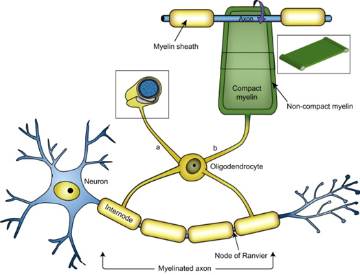
But a frequent, insightful commenter called “mauraelizabeth3” asked this question after reading my last post about a new genetic finding in PSP incriminating to the myelin-producing cells, the oligodendrocytes, as a major possible starting point for PSP.

Is it known how these findings relate to (or perhaps result in) the hyperphosphorylated tau protein that aggregates in the brains of PSP patients?

I’ll respond to that excellent question with my own current theory of the etiology and pathogenesis of PSP.  It’s based on legit science, but of course, I don’t really know how much emphasis to place on each of the disparate current facts, or how many additional facts await discovery.

Dear me3: 

That’s really the question, isn’t it!  My own formulation at this point is that the loss of myelin from those multiple gene variants is a sideshow that impairs neurological function but isn’t actually part of the cause of PSP.  Instead . . .

  1. I’d propose that the first abnormal event is some inherited or de novo mutations or epigenetic alterations in the MAPT gene (which we know do exist in PSP) changes the structure or the post-translational modifications of tau in a way that stimulates its hyperphosphorylation as a reaction designed to facilitate its degradation.  Hyperphosphorylation tau might also be the result of some sort of toxin exposure, with metals currently the leading contender.
  2. Then, hyperphosphorylated tau falls off the microtubules and is free to do mischief all over the cell.  Maybe the first (or only) thing it does is to make the genomic DNA in the nucleus lose some of its protection against inappropriate transcription into RNA.  It’s been shown that such inappropriate transcription allows retrotransposons to be transcribed.  (Those are pieces of DNA implanted there by viruses millions of years ago.  They have reproduced themselves to other parts of the genome and now account for about 40% of our DNA.)  
  3. The RNA so produced is recognized by the immune system as viral.  An immune response ensues, which attacks and degrades a lot of RNA and innocent bystanders.  The resulting molecular garbage is a major challenge for the cell’s regular garbage disposal, the ubiquitin-proteasome system and the autophagy/lysosomal system. 
  4. Meanwhile, all that hyperphosphorylated tau is misfolding and then aggregating with itself.  This does take place under normal, healthy conditions to some extent, and the disposal systems can handle that.  But now, with the garbage from the inflammation and the unusual amount of aggregated tau to degrade, the garbage disposal is overwhelmed.  This allows all sorts of normal toxic garbage to accumulate, and that’s eventually fatal to the cell. 
  5. This whole process starts in the astrocytes or oligodendrocytes and spreads to the neurons courtesy of the microglia (the brain’s immune cells), synaptic connections and direct contact. 

Keep in mind that I’m a clinician with little laboratory experience beyond delivering fluid samples from my patients to my smarter colleagues with the pocket protectors.  But I try to keep up with the latest in all aspects of PSP, and there’s lab support for all of the assertions in my hypothesis.  It’s just that putting those facts together is tricky, like cracking a complicated criminal plot.  I hope that my hypothesis at least illustrates that we’re starting to get more of a handle on the pathogenesis of PSP.

Thanks for the complement

The Alzheimer’s Association’s division devoted to frontotemporal dementia and related disorders has just announced its award for best publication of the year for 2004.  It’s entitled, “Genetic, transcriptomic, histological, and biochemical analysis of progressive supranuclear palsy implicates glial activation and novel risk genes.” The lead author is Dr. Kurt Farrell of the Icahn School of Medicine at Mt. Sinai in New York, and the senior author is Dr. Adam Naj of the David Geffen School of Medicine at UCLA.  Full disclosure: I played a very minor role in an early phase of the work and am 34th of the 48 co-authors.

The study sought to identify new genes conferring risk for PSP by comparing genetic markers between a group of 2,779 people with PSP (a large majority autopsy-confirmed) and 5,584 people with no neurodegenerative conditions.  This is the largest such study in PSP to date, the first having been published in 2011. The study’s large size gives it greater power to distinguish a genuine PSP-related genetic variant from a statistical fluke. 

This technique can identify not a specific gene, but a small region of a chromosome, typically with 50 to 200 genes.  But the researchers then nominated a couple of the most likely genes in that region and tested brain tissue for excessive amounts or abnormal forms of the protein encoded by the candidate genes.

Besides confirming PSP’s previously-identified genetic risk factors, Farrell and colleagues identified a new risk gene called C4A, located on chromosome 6. The protein it encodes is part of the cell’s “innate immune system.” The C stands for “complement,” a complicated group of proteins that assist the antibodies.  Once an antibody latches onto an unwanted invader like a bacterium or virus, proteins from the complement system attach to the other end of the antibody, initiating a series of interactions that eventually produces an “attack complex” that pokes a major hole in the invader.

The other important finding from the same study was a strong tendency for all the genes known to increase PSP risk to affect the oligodendrocytes.  Those are the brain cells that form the myelin sheath insulating the axons of most of the brain’s neurons, greatly facilitating electrical conduction.  We’ve known for decades that loss of myelin is an early feature in PSP, and that some PSP risk genes encode proteins related to oligodendrocytes.  But the new paper provides important confirmation in a larger patient group, using techniques not previously available.

So, bottom line: Although the genetic basis of PSP is not usually enough to make the disease occur in more than one member of a family, it could still be one important factor, along with things like a poorly-understood toxin exposure.  Furthermore, the new evidence that the oligodendrocytes might be the first cells to get sick in PSP points researchers in that direction in their search for new, easily addressable drug targets.  Plus, the discovery that an important part of the immune system could be what’s ailing the oligodendrocytes means that the array of potential drug targets is now much better focused. Shotgun approaches to taming components of the complement system have been under way for about a decade now, and the new finding of Farrell et al could focus those efforts nicely.

An insulation gene or two

My last post reported a study demonstrating that the genetic variants (often called mutations) conferring PSP risk interact with one another to elevate the risk beyond the simple total of their individual effects. The next day brought a publication on yet another gene associated with PSP.


The current paper’s 78 authors are in Spain, Portugal and The Netherlands. The first author, Pablo García-González, is a graduate student at the International University of Catalonia in Barcelona. The senior author is Agustin Ruiz, director of the Alzheimer research center there.


The scientists analyzed 327 Iberian and 59 Dutch patients with either PSP-Richardson syndrome or with autopsy confirmation of PSP. They excluded living people with non-Richardson variants because for a significant minority of such people, the underlying condition is not actually PSP. This is the same reason most clinical PSP trials exclude people with non-Richardson variants . The advantage is less “statistical noise” from misdiagnosis. The disadvantage is that the conclusions may not apply to all variants of PSP.


The new study compares the patients and the non-PSP control subjects using 815,000 genetic markers. Variants in the markers differ with regard to which of the four nucleotides (the “letters” of the genetic code) occurs at a specific spot in the marker gene. The markers are selected because they are approximately evenly spaced along all 23 chromosomes and occur in multiple variants in the general population, a state called a polymorphism. If the frequencies of the four nucleotides at any one polymorphic location differ between the disease group and the control group to a degree unlikely to occur by chance, we call that a statistically significant allelic association. But it’s only just that – an association, and the tricky task of proving cause-and-effect begins.


The researchers confirmed previously discovered markers and found one new one nestled between two genes called NFASC and CNTN2 on chromosome 1. The proteins encoded by both genes are involved in the function of the oligodendrocytes, a major type of non-electrical brain cell. The “oligos,” as we in the business call them, produce the insulation, called myelin, around most of the axons conducting electrical impulses among the neurons

For the statistically interested: The odds ratio for the marker’s association with PSP was 0.83, with 95% confidence interval 0.78 to 0.89 and p-value 4.15 x 10⁻⁸.

The figures are from ScienceDirect.com. Above is a neuron in blue with its axon encased in myelin as insulation. The myelin occurs in sections, each of which (labeled “internode”) produced by one branch (or dendrite) from an oligodendrocyte. The green is a stylized representation of myelin starting to roll up around a section of axon.

The figures below are cross-sections of a myelinated axon as photographed by an electron microscope. The lower-power view on the left shows the axon itself with its tiny organelles surrounded by the (barely perceptible at this power) layers of myelin. In the higher-power view on the right, arrows point to the myelin layers.

The figure below shows the extent of loss of myelin from axons in the brain as imaged by three different MRI techniques listed on the left. Each column shows a different “slice” of brain. The areas shown in blue represent areas where the myelin damage in PSP exceeded that in Parkinson’s disease. In no brain area was PD worse in that regard. The scale on the right shows how the intensity of the blue represents the statistical significance of the PSP/PD difference at that anatomical point. (From Nguyen T-T et al. Frontiers in Ageing Neuroscience, 2021.)

So, at this point, what’s the evidence for a cause-and-effect relationship between these two new risk genes and PSP? One important point is that in PSP, loss of myelin and the oligos that produce and contain it are very early and important parts of the disease process. Another is that the proteins produced by the NFASC and CNTN2 genes are “co-expressed” along with two previously-discovered PSP risk genes called MOBP and SLCO1A2, both of which are also associated with oligos and myelin. Co-expression refers to a close correlation between the amounts of multiple kinds of proteins being manufactured by the cell, implying that the proteins work together to perform some specific task required by the cell at that point in time.

The discovery of these two new PSP risk genes supports the idea that the integrity of the oligos and their myelin is a very early and critical part of the PSP process. We already know a lot about that process, thanks to decades of research on multiple sclerosis, where breakdown of myelin is even more important than in PSP, but occurs for more obviously immune-related reasons. That previous research has identified many myelin-related enzymes and other kinds of molecules that might be susceptible to manipulation by drugs. Now, it’s a matter of finding the most critical such molecules and the right drugs to return their function to something like normal.