A cluster mystery

Since 2014, I’ve been trying to find the cause of a geographical cluster of PSP in northern France.  It’s the only documented PSP cluster known.  The problem was difficult enough, but now the cluster has mysteriously disappeared.

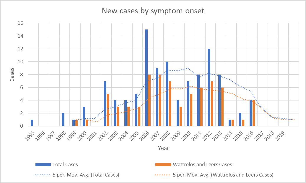
The clinical aspects of the cluster are detailed in this 2015 paper.  Here’s the executive summary: In 2005, Dominique Caparros-Lefebvre, MD, a geriatric neurologist with experience in PSP research, arrived at her new practice position in Wattrelos, France, an industrial suburb of Lille.  By 2007, she started to notice more PSP than expected and developed an excellent database.  She diagnosed 100 patients over the next decade.  In 2013, she invited me to help her find the cause, as I had had some experience in the epidemiology of PSP.  I calculated the observed-to-expected incidence ratio of PSP to be 12.3 in Wattrelos and its neighbor to the south, Leers.  Most clusters of chronic diseases such as cancer have ratios much lower, in the range of 1.5 or 2.0.  So this was a major cluster.

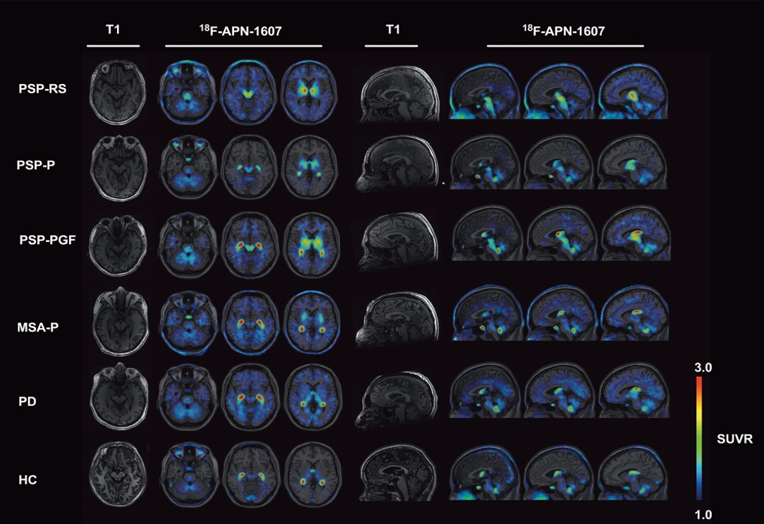
PSP in the 100 patients has differed only slightly from “sporadic PSP,” with more PSP-Parkinsonism than PSP-Richardson syndrome, and an older mean onset age, 74.3.  The 13 autopsied cases show typical PSP, with the expected 4R tau and the H1/H1 genetic haplotype.  That work was done by a very accomplished research team at the University of Lille led by Luc Bueé, PhD and Vincent Deramecourt, MD, PhD.  No other molecular genetic workup has been performed to date, but none of the affected persons were related to one another and among the patients are 7 Algerian immigrants, a strong point against a genetic origin.  

Wattrelos and Leers have extensive chemical contamination, especially by metals from an ore extraction plant that operated in southern Wattrelos for most of the 20th century.  Huge piles of spent chromate and phosphate ore, now covered, remain on the plant’s property, which has been converted into to a public park after mitigation efforts between 2000 and 2010.   Only 2 of the 100 patients worked in the chromate/phosphate ore plant, but soil from the area adjacent to the slag heaps has been used as fill in construction and road maintenance over a wide area.  Furthermore, multiple chemical-related industries such as tanning and dyeing formed the base of the town’s economy for many decades.

So, the obvious culprit has been metals.  Chromium is a carcinogen but not a good candidate as a direct neurotoxin, as its most common form, hexavalent chromium, does not cross the blood brain barrier.  Nor is phosphorus a good candidate, but phosphate ore often contains important levels of other metals. 

In France, growing one’s own herbs and vegetables is a common practice, even in densely urban areas.  Dr. Caparros suspected thyme, a widely used herb in French cooking that avidly absorbs metals from the soil.  The French government’s soil data and our analysis (by my Rutgers colleague Brian Buckley, PhD) of home-grown thyme samples from Wattrelos suggested that arsenic, cadmium and nickel were the most likely possibilities.

In 2016, I recruited a team of neuroscientists led by Aimee Kao, PhD of UCSF, with skills in stem cell models of PSP and access to stem cells with PSP-related tau gene mutations.  As an initial project, they created brain cells with a rare PSP risk mutation (to create a “background” vulnerability) and exposed them to chromium, cadmium and nickel.  They did the same experiment with cells from the same PSP patient except that the PSP risk mutation was converted to normal using CRISPR.  They found that some of the same damage seen in PSP — aggregation of tau and evidence of apoptotic (i.e., programmed cell death) in the exposed cells with the mutation.  But those abnormalities are not specific for PSP.  We published that in 2020 and unfortunately, I couldn’t keep that team together for follow-on projects.  Equally unfortunately, the local French human research authority would not allow Dr. Caparros to perform further field work that might have pointed to a specific metal and route of exposure.

So why aren’t clusters of PSP seen in the many other places in the world where those metals contaminate the environment?  My own pet theory was that toxicity from multiple metals acting in concert is needed, and no place other than Wattrelos/Leers has a combination of so many metals in one spot together with a physician able to diagnose PSP as well as Dr. Caparros.  So, one of the follow-on projects might be to repeat the lab experiments with combinations of the same and other metals that are known to occur in the environment, either in Wattrelos/Leers or elsewhere, either as a result of industrial pollution or naturally occurring.

As I was starting to make plans for such a project with lab colleagues at Rutgers, it became clear that the number of patients whose onset occurred since 2013 has been declining.  The most recent onset date of the 100 patients is 2016.  This is not how a cluster of toxic (or genetic) cause should act.  It’s possible that the mitigation efforts on the two slag heaps reduced the rate of entry of chromium and phosphate into the local environment, but the metals were pervasive in the area and presumably remain so.

—-

Graph showing the disappearance of cases with onset since 2016. Bars show the number of cases with onset in each year. Dotted lines show the average over the previous 5 years. “Total Cases” refers to all cases in Wattrelos and Leers as well as in nearby towns where patients were likely to use the Wattrelos hospital. The paucity of cases with onset before 2002 is explained by the arrival of Dr. Caparros in Wattrelos in 2005. Before that, no physician likely to have been able to diagnose PSP worked there and few patients with onset before 2002 would have survived to come to Dr. Caparros’ attention. Still, we cannot rule out the possibility that the cluster in fact started in the 1990s and its disappearance by 2017 is consistent with this possibility.

—-

The unexplained abatement of a geographical or temporal disease cluster speaks for an infectious cause.  The salient example of an infection causing a temporal cluster of a neurodegenerative tauopathy is postencephalitic parkinsonism (PEP).  That cluster started 2 years before and ended a decade after the great “Spanish” flu pandemic of 1918-1920 and is independent from it.  PEP was a chronic, levodopa-responsive parkinsonism that affected people of any age who recovered from an encephalitis that was presumably viral, but the specific virus has not been identified.  At autopsy, the brain showed neurofibrillary tangles not very different from those of PSP.  The last patients with PEP died before modern molecular techniques were available, so its cause may never be known.

Could the cause of the Wattrelos/Leers cluster have been a virus?  True, there seemed to have been no antecedent encephalitis, but it may have been mild, self-diagnosed as a cold or the flu, and forgotten by the time Dr. Caparros saw the patient decades later.  But there need not have been any clear acute-phase symptoms at all.  The virus could have set up a slow process of damage involving tau aggregation, starting with inserting its own genetic material into that of the host.  Or the initial infection could have altered the patient’s immune system in a way that encouraged (or allowed) the pathology of PSP to develop.  Let’s not forget that disordered immune modulation is one of the up-and-coming theories of PSP-causative factors.

If a virus contributed importantly to the cluster, could ordinary, “sporadic” PSP outside of the cluster be the result of a similar virus?  Or maybe sporadic PSP is caused by the same virus without the predisposing local factor of the unusual metals exposure.  Or maybe a virus infected the gut microbiome of the Wattrelos population in a way that increased PSP risk.  I could go on.

We know that at least one neurodegenerative condition, sporadic Creutzfeldt-Jakob disease, is caused by an infectious agent (in this case the prion protein) without geographical or temporal clustering.  The idea of a virus or prion as a cause of PSP is not new, and previous attempts to prove that hypothesis starting in the 1970s have been negative.  But the technology for finding viral fingerprints has improved markedly since then.

I’ll try to get some of the research honchos I know interested in this theory and get back to you.

A warning

Here’s something that may seem too good to be true, and in my opinion, it is.  I’m writing about it as a cautionary measure.

Researchers at Guangzhou University, China have published a case report of a man with advanced PSP who received intravenous and intrathecal (into the spinal fluid) infusions of stem cells derived from umbilical cord blood.  His symptoms had started unusually young, at age 53, and 8 years later, his PSP Rating Scale score was 73 – a typical score for that duration of disease.  He received the infusions at that point and has survived another 8 years to date with essentially the same PSPRS score. 

Although the patient is (apparently, but not explicitly) still alive and there has been no autopsy to prove the accuracy of the diagnosis, the clinical history, neuro exam and published MRI images are typical for PSP.  The subtype is probably PSP-Parkinson, as his falls didn’t start until 3 years in and he didn’t need gait aids until 2 years after that.  The authors state that the subtype was PSP-Richardson syndrome because the patient met those criteria at the time they first saw him, 8 years in.  But PSP-P usually evolves into PSP-RS in the advanced stages.  The life expectancy of PSP-P with typical onset age in the 60s is about 10 years, and for those with onset as young as age 53, would be a few years more.  So survival (to this point) of 16 years is not far from expected.

But that’s not the main problem. The main problem is that the PSP Rating Scale has a “ceiling effect.”  That is, patients progress at an average rate of 10 or 11 points per year, and once the score reaches the 70s, it stops progressing although the patients continue to survive and to worsen.  That’s because the worst possible score on many of the scale’s 28 items doesn’t capture the full level of dysfunction that can yet occur.  Another reason for the ceiling effect is that some of the 28 items don’t affect some patients severely or at all, even late in the disease course.  Examples are dystonia, tremor, irritability, emotional incontinence, horizontal eye movement loss, and limb apraxia.  So the patient of Dr. Li et al may simply have reached his PSPRS ceiling and continued to survive by virtue of the unusually good general care that study subjects typically receive.

There are some other issues:

  • There’s no objective evidence that the infused cells survived. 
  • There’s no measure of any sort of growth factor in the blood or CSF that might provide a mechanism of action of stem cells in halting an otherwise rapidly progressive disease in its tracks. 
  • There’s no functional measure of brain metabolism such as fluorodeoxyglucose PET or magnetic resonance spectroscopy to objectively document a halting/slowing of progression.
  • The 2 MRI were performed at the time of the infusions and 2 years later.  Although the authors claim “no deterioration” in the MRI over that time, the 2 sets of images do show progression of atrophy, both to my eye and by the formal measurements superimposed on the images.  The midbrain’s diameter on the before-and-after sagittal images (Fig. 1, images A-1 and B-1) declined from 11.84 mm to 10.64 mm and the pons from 21.07 mm to 19.37 mm.  Both are typical for PSP.  The before-and-after axial images (images A-2, A-3, B-2 and B-3), where the measurements seem to indicate some improvement in the atrophy, are performed different scanning planes.  That can seriously affect the simple measurements performed. In fact, comparing the 2 sagittal images shows that the patient’s head is at 2 very different tilt angles in the scanner.  That problem affects the measurements in the axial plane but not in the sagittal plane.

As Dr. Li and colleagues point out, “randomized controlled trials are needed in the future . . . “ I would advise people with PSP not to undergo this, or any, experimental procedure outside of a formal trial at a reputable academic institution.  If you’re considering it, make sure the study is listed in clinicaltrials.gov, although that by itself no guarantee of anything.  Make sure that the researchers have a track record of peer-reviewed publication in this area.  If the doctors doing the treatment are willing to use it for a wide variety of unrelated disorders, be suspicious.  If the claims omit mention of side effects or toxicity, be doubly suspicious. If there’s no mention of success in any sort of animal model, that could be a problem.  Finally, if a “research study” charges a hefty fee, stay away. (We have no evidence that this was the case for Li et al.)

We should encourage fresh ideas for the treatment of PSP and other neurodegenerative diseases.  We should not be biased against research from countries with still-developing research infrastructure and institutional safeguards.  But we should also know how to evaluate the quality of research reports and to be vigilant for signs of quackery.

New info we can use this afternoon

If you like my research updates but need a break from the molecular stuff, here’s an interesting and unexpected clinical discovery, usable at routine visits for care of PSP.

Michelle Troche, PhD, is a speech/language specialist in the Laboratory for the Study of Upper Airway Dysfunction in the Department of Biobehavioral Sciences at Columbia University’s Teachers College.  She’s a past grantee of CurePSP’s

for a project published a year ago on training patients with PSP to protect their airways from aspiration. Now, she and her team, with James Borders as first author, have analyzed cough function in quantitative detail in patients with PSP and Parkinson’s.

The two groups of 26 patients were designed to be similar in terms of age, sex, disease duration and severity of swallowing difficulty.  Their cough was analyzed with a device called a pneumotachograph (“air-speed-writer’) that fed the data into a software system.  The procedure was performed during both coughing on request and coughing induced by a 2-second spray of capsaicin in 4 progressively increasing concentrations.  Capsaicin is an irritant found in hot peppers, so kudos to those volunteer patients!

After a sophisticated statistical correction for confounding factors, some results were as expected: that the patients with PSP were able to generate only a fraction of the expiratory flow rate and volume of those with PD.  However, another result was unexpected: patients with PSP were more bothered by increasing concentrations of the capsaicin than were those with PD, but were no more likely to respond by coughing. 

In the researchers’ words, “. . . it is interesting to note that although both groups exhibited blunted urge-to-cough slopes compared to prior research in healthy adults, patients with PSP demonstrated increased urge to cough compared to PD. This means that even though the participants with PSP were perceiving the increasing cough stimulus more than patients with PD, they were not coughing more to that stimulus.

So what does this mean?  We think of PSP as a motor and cognitive disorder, but this result shows that there’s also a sensory deficit.  The sensory input into the cough reflex takes place in the brainstem, the main location of pathology in PSP, so this result makes sense.  It’s just that no one had previously thought about it. (The same phenomenon accompanies many important scientific discoveries – they seem so obvious in retrospect.)

Another brainstem reflex, the auditory startle response, has long been known to be impaired in PSP but not in PD, so the results of Borders et al have that ex ante confirmation.

Like any pioneering work, this one has its limitations.  Although the PSP and PD groups were similar in terms of disease duration (both about 5 years) and swallowing function, the PSP group was much worse in terms of overall disability as measured by the widely-accepted Schwab and England Activities of Daily Living (SEADL) scale. With 100 being normal on the SEADL, the PSP group averaged 48, the PD group 79.  This is expected given PSP’s more rapid disease course from onset to death — the 5-year disease duration is a far greater fraction of the average survival in PSP (7 years) than in PD (15 years).  So perhaps the authors should/can add the SEADL score to their statistical model.  Another issue is that the experimental procedure did not consider any effect on the cough function of PD medication, which dramatically helps general motor function of PD but not of PSP.

Now, how can we use this information?  Dr. Troche and colleagues suggest that patients with PSP be monitored closely for cough deficit and that their own, previously published protocol for sensorimotor training in PSP, referred to above, could be instituted sooner rather than later.

Short stuff

Most of my posts are long — maybe too long. The charitable explanation is that I can’t resist my instincts as a professor to explain stuff so my learners can understand it. The less charitable explanation is that I’m just a windbag. So here are a bunch of very brief items of news, ideas and opinion about PSP and CBD in the style of Twitter. In fact, I’ll even limit my character count to 280, including spaces. Here goes.


A group in Bologna did skin biopsies to look for a phosphorylated form of α-synuclein in PD, PSP or CBD, and controls – 26 subjects in each group. They found it in all 26 with PD, in no controls, and in 24 with PSP/CBD. (The other two had PD-like features.) Now: how about MSA?

You’ve noticed that CurePSP’s publicity materials call PSP, CBD and MSA “prime of life” diseases because those conditions’ usual decades, the 50s, 60s and 70s, are when life can otherwise be lived to the fullest. Do you agree? Let me know.


A group called the PSP Research Roundtable was formed in 2017 to help speed the process of testing promising drugs. It’s run by CurePSP and has membership from academia, the FDA, the NIH, drug companies, biotech, philanthropy and patient advocacy groups.


Transposon Therapeutics has started a Phase 2 trial of TPN-101 in PSP at private clinical trial sites in Boca Raton, FL and Farmington Hills, MI. Like many available HIV drugs, TPN-101 inhibits the enzyme reverse transcriptase, but otherwise, details are sparse.

About the mechanism of action of TPN-101: I can tell you that another reverse transcriptase inhibitor routinely used for HIV called efavirenz (trade names Sustiva and Stocrin) reduces tau aggregation. CurePSP is currently supporting a study of it in a mouse tauopathy model in The Netherlands.

Enough for now. No windbag, I.

Marker development: anarchy vs plutocracy?

You’ve heard me whining that we need a diagnostic “trait marker” for PSP. In other words, we need to be able to accurately distinguish PSP – during life — from such mimics as Parkinson’s, multiple system atrophy, Alzheimer’s, corticobasal degeneration, normal-pressure hydrocephalus and others. Only in that way can we create “pure” groups of patients in which to study the disease and test specific treatments.


Right now, the best diagnostic test we have is the MDS-PSP Diagnostic Criteria, which requires only traditional history-taking and a hands-on neurological exam. Those criteria work well for PSP-Richardson syndrome after the first couple of years but not quite well enough for earlier-stage PSP-RS nor for the “minority” or “atypical” types, which together account for 60 to 75 percent of PSP.


The most promising markers using laboratory or imaging data are levels of phosphorylated tau and neurofilament light chain (NfL) in the spinal fluid and blood; and perhaps MRI measurements of the size of the midbrain, pons and related areas at the base of the brain. But these are far from ready for prime time. NfL is a protein component of brain cells that has been shown to occur at about a two-fold higher level, and to increase faster over time, in PSP and MSA than in PD, Alzheimer’s and other neurodegenerative diseases. MRI changes don’t occur in the early stages. Positron emission tomography is coming along, but won’t be ready for use in PSP for another few years, and even then will be costly and not widely available.


Last week, my routine surveillance of new PSP-related research papers in the literature yielded two interesting hits — both about PSP trait markers, both using new lab techniques, and both from Italy.

Corinne Quadalti and colleagues at the University of Bologna measured NfL and alpha-synuclein in spinal fluid and blood. They found that plasma NfL alone worked very well in distinguishing PD from PSP, with an accuracy of 0.94. (“Accuracy” in this context is the area under the receiver operating characteristic curve, which compares sensitivity with specificity. Perfect accuracy is 1.0 and a useless test’s accuracy is 0.5, where a coin flip would work as well.)


Alpha-synuclein is the main protein aggregating in Parkinson’s, dementia with Lewy bodies and multiple system atrophy. It is to those diseases what tau is to PSP and CBD. To measure it, they used a new technique called “real-time quaking-induced conversion” (RT-QuIC; pronounced, “R-T quick”), which measures that protein in its misfolded and aggregated forms. This prevents that abundant protein in its normal form from swamping the measurement. The result was positive in 91% of their patients with PD and in none of their 58 patients with PSP or CBD.


Now, if you have a nose for statistics, you’ll raise your hand and say, “But those 9% of PD patients with a negative test comprise more people in the general population than all the patients with PSP or CBD, so a negative test doesn’t mean much.” and you’d be right. So, while the sensitivity of the test for PD is excellent, the specificity is low, rendering the overall accuracy in a real-world situation insufficient.


For that reason, the authors combined two measurements – spinal fluid NfL and serum alpha-synuclein, with a resulting improvement in distinguishing PD from PSP/CBD to a sensitivity of 97.4% and specificity of 100%. That’s more like it, but keep in mind a few issues: They combined PSP and CBD into one group, and we don’t know if the results apply as well to each disease alone. They had no autopsy confirmation of the diagnoses, which means that these patients were already at a stage that was possible to diagnose using traditional clinical criteria; this means that patients with earlier-stage illness will be needed in a follow-up study. Finally, and as always, the results have to be confirmed at other centers using other techniques.


The other eye-catching paper was from Ida Manna and colleagues at the University Magna Graecia in Catanzaro, Italy. They use exosomal micro-RNA (miRNA) in blood to distinguish among PSP, PD and healthy controls.

Exosomes are tiny bubbles of brain cell membrane enclosing whatever cell contents were there when the bubble pinched itself off and floated free. They often find their way into the bloodstream. MicroRNAs are stretches of RNA averaging only about 22 nucleotides. They do not encode proteins as messenger RNA (mRNA) does, but instead bind to mRNAs to regulate their translation into protein. They are specifically encoded in the DNA of the genome and about 2,000 of them are known to exist.


Dr. Manna et al measured levels of 188 miRNAs for which there is evidence of association with some neurodegenerative disease. They found a set of 6 miRNAs that together yielded an accuracy in distinguishing PSP from PD of 0.91. The accuracy for distinguishing PSP from controls was 0.90.
Of course, many of the same caveats that I listed for the other paper apply to this one. Plus, PSP mimics other than PD were not included in the analysis. Just as important is that there were only 25 patients with PSP and they were a mixed bag of 20 with PSP-Richardson and 5 with PSP-Parkinson. In applying a marker for the purpose of excluding patients with PD from a study of PSP, it is critical to be able to distinguish PD from PSP-P. It is unlikely that those 5 patients with PSP-P constituted a statistically valid sample for that purpose. That will be a project for another day.


What do I take away from these two papers? Neither of them alone provides a marker just yet, and each has its drawbacks given the current early stage of work. But perhaps, with some refinement, combining them with other non-invasive markers could create a diagnostic panel with enough accuracy to distinguish PSP from all of its mimics. After all, in medicine in general, multiple diagnostic tests (several tests of body fluids, some imaging, a physiologic test such as an EKG) must be combined to produce an actionable diagnosis. Why should PSP be any different?


I think the problem (and it’s a good problem to have) is that new candidate markers are being identified all the time, as are ever more sophisticated technology for measuring them, with RT-QuIC, miRNA and exosomes as prime examples. That means as researchers turn their attention to early-phase development of newer ideas using newer technology, ideas that looked potentially useful if pursued further may be neglected and not developed into practical tests. What to do? Do we just let scientific nature take its course in its traditional, anarchical way, waiting for research groups to take techniques with good initial data to the next level? Or should a group of experts with an iron fist issue some sort of “white paper” listing which markers with good preliminary evidence, perhaps like the ones I describe here, should be nurtured with funding and collaborations? If so, who chooses those experts? And once the experts are chosen, how can we prevent them from favoring the ideas in which they’ve invested their own time, resources and reputations?


You know where the “comment” button is.

An RNA surprise

As usual, some background may be helpful here: You’ve heard of genomics, the analysis of large numbers of gene variants as a clue to disease causation.  But genomics doesn’t deal with the actual proteins produced by those genes.  That led to proteomics, the analysis of proteins in a tissue or fluid sample, as a more relevant window into disease mechanisms and possibly as a path to diagnostic tests.  But proteins in samples can be influenced by many things such as breakdown of proteins by enzymes or by the cells’ normal garbage disposal mechanisms.  A solution is “transcriptomics,” which identifies and quantifies messenger RNA (mRNA) on its way from being encoded (“transcribed”) by DNA to being translated into protein.  Although every cell in our bodies has our entire genetic endowment in the form of DNA, only a fraction of those genes is actually transcribed into mRNA.  The types and amounts of mRNA produced depend on the job of the specific cell and its protein needs at the moment.  Click here if you’d like to chew on a highly technical but excellent, current review of the topic.

In the current issue of the prestigious Journal of Clinical Investigation, a team mostly from the Mayo Clinic Jacksonville has compared the transcriptomes of tissue from cerebrum and cerebellum with autopsy-confirmed PSP to those with Alzheimer’s disease and to controls with no autopsy evidence of neurodegenerative disease.  I’ll cut to the chase:  To the researchers’ surprise, they found the four types of tissue to be quite similar in their transcriptomic profiles.  This result suggests that a treatment or a diagnostic test directed at one or more of the protein (or mRNA!) abnormalities in either disease could work for the other disease as well.  We already suspected that, but without a lot of supporting evidence beyond the superficial observation that both diseases involve tau aggregation.  This new paper provides some better evidence.

I’ll return to the paper’s practical implications in a minute.  But first, back to biology class you go: “O-omics” results (Wikipedia lists 45 kinds of “omics” in biomedicine to date) are by their nature shotgun approaches, typically yielding a long, inscrutable list of statistically significant but questionably meaningful differences between subject groups.  So, seeking some sort of useful pattern in the data, researchers divide the genes, proteins or mRNAs yielding statistically significant “hits” into categories based on their known functions.  These categories are called “networks” because they’re based on interactions or on commonalities of function.  The process of creating and using such standardized, defined categories in genomics is called “gene ontology.”

The authors of the new paper point out that previous transcriptomic work in AD has revealed differences from controls in many such networks, most prominently “immune function, myelination, synaptic transmission and lipid metabolism.”  They also point out that it’s usually difficult to know if these mRNA differences are the cause of the cellular damage in AD or merely the brain’s reactions to that damage.

Now back to this project:  As I said above, the transcriptomic work analyzed not only the cerebral cortex, where the researchers knew ample pathology exists in both AD and PSP, but also, as a kind of control group, the cerebellar cortex, where the standard autopsy shows little or no damage in AD or PSP.  Another feature of the study design was to use the temporal lobe as the source of its cerebral samples.  That part of the brain is heavily involved in AD by standard methods but little or not at all in PSP. 

Surprisingly, the results were that the transcriptome abnormalities (relative to non-neurological controls) were quite similar in all four types of tissue – AD temporal cortex, PSP temporal cortex, AD cerebellar cortex and PSP cerebellar cortex.  In their words, “The DEG [differentially expressed gene] changes between AD and PSP in two regions of the brain demonstrate a striking conservation [consistency] of transcriptomic changes across these different neurodegenerative diseases.”

Because the degree of traditionally measurable cell damage differed markedly across those four sets of samples, they infer that the changes are “upstream,” meaning at an early step in the disease process, rather than “downstream,” in reaction to damage.  That would mean that even in the early stages of AD and PSP, the disease process is already at work in areas that have not started to show physical signs of damage as assessed by microscope, MRI or PET scan.

What were the networks most affected by the transcriptomic changes?  In the authors’ words: “Up-regulated in both AD and PSP were gene networks serving chromatin modification, gene expression, chromosome organization and metabolism of nucleotides. In the cerebellum the shared upregulated genes link to biological processes relating to RNA and RNA transcription, cell-cell junctions, and heart, kidney, gland, and circulatory system development. Shared down-regulated genes in AD and PSP are associated with gene ontology cell compartment terms related to mitochondrial and ribosomal functions in both the temporal cortex and the cerebellum.”  

The paper’s first author is Xue Wang, PhD, a bioinformatician at Mayo.  The last two authors, sharing credit as senior authors, are Todd Golde, MD PhD of the University of Florida and Nilufer Ertekin-Taner, MD PhD.  I emailed Dr. Taner for her take on the results.  She replied in part, “These findings can be leveraged to develop multifaceted therapies and biomarkers that address these common, complex and ubiquitous molecular alterations in neurodegenerative diseases.”  I’d agree.

So, this unexpected discovery suggests that it may prove fruitless to look for causes of specific neurodegenerative diseases in their gene expression profiles.  Abnormal gene expression may not be the true origin of the disease, but only the cell’s reaction – not to downstream physical damage visible with standard tools, but to some other, far more upstream causation.  Yet, interrupting that reaction at the level of its mRNA might be the key to halting the progression of multiple, of not all, neurodegenerative diseases.  Adding to the appeal of that approach is that the treatment targets are available at a very early stage of the disease process.

Testing that suggestion will have to start with analyzing more than just AD and PSP, and I’m guessing that this effort is already in progress thanks to the excellent collection at the Mayo Brain Bank.  I’ll keep you informed.

A shock to the system

Researchers at McGill University in Montreal have reported improvement in gait speed in a woman with PSP using transcranial direct current stimulation (tDCS). 

The research findings that I pass along here are generally of high scientific quality.  This one is only a single case report and was published only as a letter to the editor, which typically meets a lower standard in the peer-review process.  But it’s in a good journal and from a well-reputed group with a long record of accomplishment in a closely related field.  Plus, it’s about a low-risk potential treatment of PSP — a disease without much other treatment.  So – good enough for me.

The paper’s first author is Carlos Roncero, MD, PhD, a psychiatrist and psychologist and the senior (i.e., last-named) author is Howard Chertkow, MD.  Both McGill professors have long and distinguished research records.  Dr. Chertkow has worked extensively on tDCS for Alzheimer’s disease and is perhaps most famous for having developed, along with two colleagues, the Montreal Cognitive Assessment (MoCA), a quick test of general cognition that works very well in PSP and other frontal lobe conditions and is used world-wide. 

There has been research before in both tDCS and in transcranial magnetic stimulation for movement disorders, including a bit in PSP.  But the previous work has used arcane physiological variables or speech as their outcome measures rather than gait or balance.

The methods

The procedure consists of passing a weak electric current through the brain, in this case from two electrically negative electrodes (“anodes”) on the skin, one over each deltoid muscle (at the shoulder), to a single positive electrode (“cathode”) atop the center of the head, where the left and right “primary motor cortices” nearly touch.  Nothing pierced the skin – these were just wires ending in 5 x 7-cm (2 x 3- inch) patches held by adhesive. Each deltoid received 2 milliamps of current over 20 minutes per day for 4 consecutive days per week for 3 weeks.  The patient’s gait was tested during the fourth stimulation of the third week and then monthly for 5 months after the stimulation sessions had ended.  As a placebo control, before the first week of stimulation, they gave the patient a week of sham stimulation, with the apparatus in place but the switch off, and considered the gait result at that point to be her baseline.  The nature of the gait test was the time required to walk 24 meters (26 yards) using the same walker that the patient was using at home.  Three clinicians flanked her to prevent falls but did not touch her or the walker.

Here are the results:

Note that the “interval time” on the vertical axis is the time to cover 3 meters, calculated by timing the patient for the 24 meters and dividing by 8.  The average healthy woman of that age (61) walking at maximum speed covers 3 meters in about 1.6 seconds and walking comfortably in 2.3 seconds. (I calculated those times from the reference data in this publication.)  With only sham stimulation, the patient’s time for 3 meters was 11.92 seconds.  It sped up to 9.46 seconds by the end of the third week. The time improved further a month later and further still a month after that, to 7.47 seconds.  After another month, it started to return to baseline and returned a bit further a month later, but then stabilized at about 9.8 seconds for 3 weeks.  So they resumed the stimulation and the next week brought improvement to 8.72 seconds. 

There are some methodological issues. 

Unfortunately, the gait was not tested pre-sham and the patient was not asked if she knew that she had received a sham treatment and when it was given.  If the real stimulation had produced a bit of an electrical sensation in her skin, that could have had a placebo effect with a resulting false-positive result.

Secondly, we don’t know how much, if at all, the speed would have improved beyond that 8.72 seconds if after 5 months had they had given the treatment for the same 3 weeks as the first time.  We also don’t know if this degree of improvement made a difference to the patient’s activities of daily living; nor if it was accompanied by an increased risk of falling not observable over the short time sample of the tests. 

Another caveat is that the gait was assessed using a simple timing of gait speed with a walker rather than with an automated gait analysis system.  Such devices are available commercially and typically have the patient placed in a harness to prevent falls and monitor dozens of variables transduced through electronic contacts embedded in a long walking mat. 

The clinicians with the patient during her walking tests were aware of whether she was receiving sham or real treatment and could have unconsciously influenced her performance. 

Bottom line

In summary, this harmless electrical stimulation procedure may eventually prove to give moderate improvement in gait speed in people with PSP, with long-term retention of benefit.  This result could serve as justification for a grant to study the issue in a larger group of patients and using more rigorous procedures and an assessment of improvement in the patients’ activities of daily living.

The methodologic informalities I’ve complained about are standard in exploratory research, which I’m sure is why this prestigious journal’s editor accepted the manuscript.  This is a good example of how science doesn’t just come up with new knowledge, “eureka!”-style.  The process is full of fits and starts, blind alleys, disagreements, human error, and lots of sweat, with one piece providing a toehold for the next until something useful emerges and is confirmed by others.   

OK, so maybe we do have a marker.

You may recall a post from last week lamenting the state of diagnostic markers for PSP.  But now I’m happy to report that things are starting to look up. 

A paper in the current issue of Movement Disorders is from a group at Fudan University in Shanghai led by Dr. Ling Li.  Two of the 17 authors work at Taiwan-based Aprinoia Therapeutics.  Last on the author list is the “Progressive Supranuclear Palsy Neuroimage Initiative” (not to be confused with the 4-R Tau Neuroimaging Initiative based at UCSF under Adam Boxer).  I don’t know if the PSPNI is an academically-based research group or a consortium created by Aprinoia.  I’ll try to find out.  In any case, Aprinoia is developing a PET tau ligand called [18F]-APN-1607, formerly known as [18F]-PM-PBB3. 

First, a little background:

What’s a “PET ligand”? What’s “PET”? Positron emission tomography is a way of mapping the locations of a specific compound (called the “target”, typically a protein of some sort) in the body. First, a compound (the “ligand”) that can bind to the target, and hopefully only to that target, is formulated, and that’s the hard part scientifically. Then the ligand is attached to an atom that emits radiation, specifically positrons, for a time that’s short enough to avoid poisoning the patient or the environment. The most common positron-emitting atom is fluorine-18, but carbon-11 is another common one you’ll see. The resulting compound is injected intravenously into the patient. In about an hour or so, the ligand has bound to its target molecule. After a positron has traveled about a millimeter, it has lost enough energy that when it next hits an electron, the two annihilate each other, emitting two photons (in this case also called gamma particles) in opposite directions. The patient is precisely positioned next to a type of camera that can detect these, and when it detects two photons at exactly the same time, it calculates their common point of origin and puts a dot on its software map accordingly. The result is a series of 2-dimensional slices showing the locations of the positron emitter with its ligand. PET images are initially just shades of gray but for ease of eyeball interpretation are typically displayed in an arbitrarily chosen array of colors, with the “cool” blue colors signifying low ligand uptake and “hot” reds the highest uptake.

Although the FDA approved Tauvid (flortaucipir; [18F]-AV-1451; [18F]T807) in May 2020 as a tau-directed PET ligand for Alzheimer’s, neither that compound nor several other candidates have proven adequate in PSP.  The main reasons have been that the “tau burden” in PSP is only 1% of that in AD, which makes the PET signal insufficiently distinguishable from the normal brain’s background.  Also, PET in general has a much lower spatial resolution than MRI or even CT, so the small size of PSP’s specific areas of involvement makes it hard for PET to distinguish PSP from other disorders.  Another issue has been non-specific binding. That is, some candidate tau PET ligands bind less to tau than to other compounds that tend to occur in the same set of brain cells but may not be affected much in PSP.  A good example has been [18F-THK-5351, which distinguishes PSP from healthy people, but was found to bind mostly to monoamine oxidase B, an enzyme important in dopamine metabolism.

Another ligand,[18F]-PI-2620, has avoided that pitfall and distinguishes PSP from healthy controls.  But it has not yet been shown to distinguish PSP from other atypical parkinsonisms, though adequate studies of that question have not been published.  Nor has [18F]-PI-2620 been tested in patients with early PSP, where there is greatest need for a diagnostic marker – the average PSPRS score of the patients in the one published diagnostic study was 38 (0 normal, 100 worst possible), by which time PSP is usually easily diagnosable at the “bedside.” (https://www.ncbi.nlm.nih.gov/pmc/articles/PMC7341407/)  Nor has that ligand been tested for its ability to distingish PSP-RS from other subtypes or to track disease progression over time.

This week’s development

The news flash is that [18F]-APN-1607, has leapt ahead of [18F]-PI-2620, at least for now.  (Not that we shouldn’t have multiple tau PET ligands for PSP with slightly different properties for different clinical situations – that would be great!)  The paper of Li et al included 20 patients with PSP (a lot for an early-phase PET study), of whom 16 had probable PSP-Richardson syndrome, 2 had PSP-parkinsonism, 1 had PSP-progressive gait freezing and 1 had “suggestive of” PSP.  Their average PSP Rating Scale score was 31.6, which is toward the milder end of the range typical of PSP drug trials and milder than the patients in the l[18F]-PI-2620 trial.  There were also 7 with MSA-parkinsonism, 10 with Parkinson’s disease (both of which are alpha-synucleinopathies, not tauopathies) and 13 healthy controls.  The results were corrected for any effects of age, sex, or disease duration and for multiple comparisons.   

The study found that [18F]-APN-1607 PET shows major differences between PSP and healthy controls in 12 brain regions known from autopsy studies to be affected most in PSP. The same could be said for the comparisons of PSP with Parkinson’s or MSA-P, although when only the putamen (part of the basal ganglia) was considered, 4 of the 7 patients with MSA-P had as much binding as those with PSP.  So the authors combined the measurements from the substantia nigra (part of the midbrain, which is part of the brainstem) with those of the putamen, achieving much better separation.  Still it was far from perfect: The standard measure of diagnostic accuracy at an individual patient level, as opposed to merely comparing two groups’ average measurements, is the area under the receiver operating curve (AUC).  That statistic, where perfect is 1.0 and useless is 0.5, takes into account both sensitivity and specificity.  The AUC based on [18F]-PI-2620 uptake in putamen and midbrain for PSP vs the synucleinopathies was 0.811 and for PSP vs. controls, 0.909.  Good but not great.

When they homed in on the subthalamic nucleus, a tiny area that may be where PSP starts in the brain, the AUC was an excellent 0.935 (0.975 for MSA-P and 0.908 for PD).  But that nucleus is so small relative to the spatial resolution of PET that it could be a problem to train large numbers of radiologists and technicians to measure it in the real world using real-world hardware and software.

Li L, Liu F-T, Li M, et al. Movement Disorders 36: 2314-2323, 2021.

In the figure above, the first and fifth columns are the MRI images used as templates on which the PET images (the colored areas in the other columns) are superimposed. The group of images on the left are axial images through the planes of (from left to right) the pons, midbrain and putamen. On the right are sagittal images through planes a bit left of midline, midline and a bit right of midline. Each row is one patient with the condition listed at the far left. (HC means healthy control.) Note that all three subtypes of PSP show strong uptake of the tracer in the putamen and midbrain and none of the other patients shows this combination. The brain area with the greatest difference between PSP and non-PSP, the subthalamic nucleus, is too small to appear to the naked eye as a clear and separate dot in these images.

Flies in the ointment

A major pitfall for [18F]-PI-2620 is its sensitivity to light, which renders it inactive.  A solution to this problem would require not only opaque containers, but also opaque IV tubing.  This can be achieved by wrapping transparent tubing in foil, a standard procedure in hospitals for other photosensitive drugs, but one with obvious drawbacks.

The study of Ling et al did show, for several brain regions, a weak correlation of PSPRS score with [18F]-PI-2620 uptake.  The association was best for the raphe nuclei, an area of the pons (in the brainstem) with widespread connections that use serotonin as their neurotransmitter and are most closely associated with control of sleep.  Weaker, but still statistically significant associations were found also for 5 other areas.  Another selling point for [18F]-PI-2620 is that the PET signal did not correlate with the subject’s age, suggesting that the uptake is related to the severity of the illness and not some effect of aging in the context of illness. However, the duration of illness did not correlate with [18F]-PI-2620 uptake, suggesting that this technique might not be able to document PSP progression or its slowing in response to treatment in a drug trial.

Another issue left untouched by the new publication is whether [18F]-PI-2620 can distinguish PSP from CBD.  That would require subjecting patients with corticobasal syndrome (CBS) to amyloid scanning to rule out Alzheimer’s disease as the cause of their CBS, leaving a tauopathy as the most likely, but not the only, explanation.  Nor were non-Richardson PSP subtypes evaluated, other than in those 2 patients with PSP-P. 

A possible flaw in the methodology is the relatively slow progression of disability in this group of patients (on average, 0.70 PSPRS points per month, compared with about 0.92 in other studies), suggesting some sort of atypicality (or a difference of definitions of the date of onset).  Another is that the PET measurements were obtained at one point in time, which may not have been the best point given the rate of brain uptake and metabolic breakdown of the [18F]-PI-2620.  Using a rate of uptake over time rather than an absolute maximum would have been preferable and is the current state of the art.

Ling et al emphasize that their study is only the beginning of the clinical evaluation of [18F]-PI-2620 in PSP.  Future studies should include larger numbers of patients, more non-Richardson types, CBD, and a repeat scan in each patient after 6 months or more in order to assess the ability of the technique to document disease progression in individuals.

But it’s progress!

clinicaltrials.now

My last two posts summarized the portions of the PSP Study Group’s October 4 meeting on imaging, markers and longitudinal observational studies.  This one’s on the current state of neuroprotective clinical trials.  The information is from presentations by Adam Boxer and Günter Höglinger and from informal contributions by other attendees.

First, some background

“Neuroprotective” means slowing or maybe halting the progression of the underlying disease process without improving the current symptoms or disability.  It is to be distinguished from “symptomatic” treatment, which only helps the symptoms or disability, typically transiently, while the underlying process continues. 

The four most recent failed neuroprotective treatments have been davunetide, a neurotrophic (i.e., neuron growth-promoting and repair) agent; tideglusib, a kinase inhibitor (that works by preventing abnormal attachment of phosphate groups to tau), tilavonemab and gosuranemab (both monoclonal antibodies directed against the “first,” or N-terminal, end of the tau molecule). None of these four slowed PSP progression as measured by the PSP Rating Scale or any other bedside test, although there’s controversial evidence that tideglusib slowed the progression of atrophy in relevant brain areas on MRI.  

Other hopeful PSP neuroprotective agents that have failed to work in double-blind trials in recent years.  These, in no particular order, are salsalate, an approved non-steroidal anti-inflammatory drug that reduces tau phosphorylation; TPI-287, an anti-cancer drug that improves microtubule function; coenzyme Q-10, a nutraceutical that enhances mitochondrial energy production; Juvenon, an antioxidant; pyruvate, creatine and niacinamide, other antioxidants; riluzole, a drug with multiple mechanisms that is approved for neuroprotection in ALS, where its benefit is minimal; rasagiline, an inhibitor of monoamine oxidase-B, an enzyme that produces toxic free radicals from dopamine; lithium, an approved drug in psychiatry that reduces tau phosphorylation; valproate, an approved drug in psychiatry and for epilepsy that does the same; and methylene blue, an approved drug for multiple medical problems that inhibits tau aggregation.

Monoclonal antibodies

We don’t know why the antibodies have failed to date.  Maybe tau’s the N-terminal isn’t consistently present or accessible to antibodies in whatever form of tau is relevant to the spread of PSP through the brain.  Maybe the trials started too late in the course of the disease.  Maybe not enough of the antibody was able to cross the blood brain barrier, even though the tau content of the spinal fluid as measured in the lumbar space (not near the brain) was dramatically reduced.  Maybe tau is protected from antibodies as it moves between neurons by some sort of bubble-like or bridge-like membrane structure.  Maybe the cell-to-cell transmission of tau isn’t the most critical or rate-limiting step in the pathogenesis of PSP. 

A promising bit of support for N-terminal antibody treatment comes from three patients who participated in the gosuranemab trial’s site at the University of Pennsylvania who later died and were autopsied.  Their brains showed changes in the glial cells suggesting that the antibodies had incited a clear anti-tau reaction that was absent in untreated patients with PSP.  Although the neurofibrillary tangles and other visible, insoluble tau deposits were unchanged by the antibody, the authors of the paper (and I) conclude that maybe all that’s required for clinical efficacy is some tweaking to the antibody, to its dosage, to its ability to cross the blood-brain barrier, or to the stage in the course of PSP when it’s given.               

Despite the failure of the two antibodies so far and our shortage of explanations, drug companies have continued to develop monoclonal antibodies against tau.  These are being tested (almost) exclusively in Alzheimer’s for the near future.  Zagotenemab (LY3303560, from Lilly) and semorinemab (RO7105705, from Roche) are both directed against tau’s N-terminal.  BIIB076 (from Biogen) and JNJ-63733657 (from Johnson & Johnson) are directed against tau phosphorylated at position 217.  Bepranemab (UCB0107 from UCB) and E2814 (from Eisai) target the mid-portion of tau.  Lu AF87908 (from Lundbeck) targets phosphorylated amino acid 396, near the C-terminal.  The lone PSP trial of any of these is a Phase 1b (i.e., double-blind but designed to test safety, not efficacy) trial of beprenamab at one center in Germany.  Even if the drug does well in that trial, further efforts are planned only for Alzheimer’s for the time being.

Anti-sense oligonucleotides

A Phase 1, double-blind trial of NIO752, an ASO from Novartis, is in progress at 7 sites in the US, 2 in the UK, 2 in Canada and 5 in Germany.  The 48 patients on active drug will be divided into three groups, each with a different dosage level, and 12 patients will receive placebo.  The lowest dosage level will start first and the next will start only if there is no immediate safety issue with the first. The drug must be given by intrathecal injection, which means directly into the spinal fluid by injection into the thecal sac at the base of the spine.  The procedure is identical to a diagnostic “spinal tap” except that that’s a fluid removal for diagnosis and this is a fluid administration for treatment.  This will be performed 4 times at 1-month intervals followed by another 3 months of observation.  More info is here.

ASOs are short strands of RNA with multiple mechanisms of action, each at a different step in the process of translating information from the MAPT (microtubule-associated protein tau) gene into the tau protein.  Many experts feel that this approach, being far “upstream” in the pathogenetic process, is the most promising of the current neuroprotective ideas for the tauopathies.  Obviously, the issues of safety and convenience of monthly spinal taps are potential obstacles.  ASO neuroprotection against Huntington’s disease, where the aggregating protein is “huntingtin,” was reported in June 2021 to have failed, but so little is known of the mechanisms of ASOs that this is not necessarily bad news for the tauopathies.

OGA inhibitors

To self-plagiarize from a 2015 post, a class of experimental drugs for the tauopathies “reduce tau aggregation by inhibiting OGA (O-GlcNAcase; pronounced “oh-GLIK-na-kaze”). That enzyme removes the sugar N-acetyl-beta-D-glucosamine from either serine or threonine residues [amino acids] of proteins. The opposing reaction, catalyzed by O-GlcNAc transferase, like other post-translational modifications, is a common way for cells to regulate proteins. In the case of tau, having that sugar in place reduces aggregation.”  Got all that? A major plus for the OGA inhibitors is that they, like most enzyme-inhibiting drugs, are small molecules, which means they can be taken orally.

Trials of OGA inhibitors for PSP have not yet begun and there’s no clue in the grapevine as to when that might happen.  But a first-in-human study of ASN-51 (from Asceneuron) in 40 patients with Alzheimer’s is under way in Australia. 

My sources tell me that Merck has another OGA inhibitor that has not yet started clinical testing.  It’s not even listed as a pre-clinical candidate in the latest revision of Merck’s publicly available, on-line pipeline info, which was last updated on July 27, 2021.

Although salsalate failed to slow PSP progression, another approved non-steroidal anti-inflammatory called tolfenamic acid reduces tau production. A single-center, Phase 2a trial had planned to start enrolling 24 patients with PSP at the Cleveland Clinic in Las Vegas in early 2021, but the trial start is delayed indefinitely.  The drug is available by prescription for migraine in the UK and some other countries but not in the US. 

Finally, AZP2006 (from AlzProtect) activates secretion of progranulin in the brain, reducing inflammation, and also has an independent action as a tau anti-aggregant.  It is given as an oral solution.  A Phase 1 trial in progress at three centers in France and a Phase 2 trial at the same three sites is planned.

For more technical details on neuroprotection (and symptomatic treatment) in PSP, see the excellent recent review by Lawren VandeVrede and colleagues from UCSF.

Our CurePSP Centers of Care review is mostly on symptomatic PSP treatment but includes a section on neuroprotection.

Markers: the longitudinal approach

We got plenty of candidate PSP treatments.

We got drug companies willing to risk their resources on trials for a rare disease.

We got clinical trial sites with proven records of efficiency. 

We got patients willing to make the sacrifices demanded by clinical trials. 

What ain’t we got? 

We ain’t got markers. 

(Deepest apologies to Rodgers and Hammerstein.)

Markers in this context are simply diagnostic tests, and there are two kinds – trait markers and state markers.  Trait markers allow us to distinguish people with from those without the disease, preferably in a very early stage, where treatments designed to prevent further decline would be most likely to occur and most useful to the patient.  Trait markers also allow us to exclude from a PSP trial any people who don’t actually have PSP.  State markers, on the other hand, quantify the amount of damage that’s already occurred and the degree of benefit of the experimental treatment. 

The best trait marker for PSP to date is purely clinical, meaning that it does not require any sort of imaging, automated measurements of movement, gene testing or chemical testing of body fluids.  That’s the MDS-PSP Criteria, published in 2017. Other types of tests can help exclude from consideration other conditions such as Alzheimer’s, Parkinson’s, MSA, normal-pressure hydrocephalus and vascular parkinsonism, but they are only helpful in those cases where a specific alternative diagnosis is plausible.  They don’t positively diagnose PSP; they only rule out other things.

Two up-and-coming trait markers for PSP are spinal fluid levels of tau with a phosphate group on amino acid 181 (Ptau181) and neurofilament light chain (NfL).  Ptau181 levels are below normal, on average, in all forms of PSP except for the gait-freezing type (PSP-PGF).  This contrasts with Alzheimer’s disease, where that marker is elevated, on average.  The average level of neurofilament light chain (NfL) in the spinal fluid is much higher in PSP and CBS than in controls or Alzheimer’s but is also elevated in many other neurodegenerative disorders.  So the ratio of NfL divided by Ptau181 in the spinal fluid is an good marker for PSP, but cannot distinguish it from CBD, and for PSP-Richardson, it may not be as accurate as the bedside clinical criteria. For ordinary clinical use, a blood test would be easier than a spinal tap, and the utility of these levels as a state marker has not been adequately studied, even for CSF.  That requires a longitudinal study over a period of at least a year.  So the NfL/Ptau181 ratio isn’t ready for prime time as a PSP trait marker, much less as a state marker.

The most widely used state marker for PSP is still the PSP Rating Scale, which is also purely clinical. (Disclaimer: I developed the PSPRS starting in 1995 and published it in 2005 along with my statistician colleague Pam Ohman-Strickland.)   It takes 15 minutes to administer and requires no equipment other than an armless chair, a cup of water to test swallowing — and the apparatus between the neurologist’s ears.  In recent years, modifications of the PSPRS have been shorter, easier to administer by laypersons, or more directly reflective of the patient’s daily activities.  Although all of these revisions are valid and have been shown to correlate well with the full, original PSPRS, none has been widely tested in the field, and the PSPRS remains the standard for now.  But it’s not good enough.  Its score is affected by common non-PSP conditions such as injuries, arthritis or strokes, or by PSP-related conditions; for example, orientation testing can be affected by apathy, gait testing by muscle rigidity, blepharospasm by Botox and everything by dehydration or malnutrition.  So there’s a lot of variance in the PSPRS as measured from one visit to the next.  This dictates that trials be large enough and long enough to cancel out the “statistical noise,” and that costs money.

A longitudinal study is observational – it includes no treatment.  It enrolls patients with the disease of interest, or sometimes also healthy people with histories suggesting a high risk of developing that disease.  Many longitudinal trials also enroll control subjects with no apparent risk for the disease — typically spouses, relatives or friends of those in the first two groups.  All of the subjects undergo tests at entry using whatever diagnostic procedures are being evaluated as markers, some of which are repeated periodically.  The study follows the patients through their course, at least with interim histories and physical exams.  If feasible and appropriate, autopsies are obtained to verify the diagnosis and to correlate specific autopsy features with diagnostic test results during life.  The goal is to identify which, if any, of the diagnostic tests prove able to accurately identify people with the disease in the earliest stages and which can track their subsequent course with precision.

There are presently at least 8 PSP longitudinal studies in progress: 2 in Germany and 1 each in India, Italy, Japan, Luxembourg, the US/Canada and the UK.

At the PSP Study Group meeting on October 4, James Rowe of Cambridge updated the group on the longitudinal PROSPECT-M-UK study, which is headed by Huw Morris of University College London. (“M” is for MSA, a late addition.) It now includes 21 academic clinic sites in the UK and about 700 patients, of whom about 100 have made more than the initial visit.  They have found that using MRI measures of atrophy of certain regions of the cerebrum is more precise than the PSPRS, reducing the number of patients needed for a treatment trial by nearly half.  The measures were atrophy of frontal and temporal lobes and enlargement of the lateral ventricles, an indirect sign of diffuse cerebral atrophy.  This confirms and extends the findings in the two trials of monoclonal antibodies that failed to help PSP, where MRI at the start and end of the studies provided a sharper picture of the patients’ progression than the PSPRS.  The reduction of the sample size was even more marked for CBD, but in fairness, the PSPRS was not designed for that disease.  One of the PROSPECT-M-UK study’s specimen collections is skin biopsies.  These can be used to look for tau aggregation in nerve endings, a potential early-stage, only slightly invasive trait marker.  Skin biopsies can also be used to create stem cells, which are then converted into neuronal cultures in which experimental treatments can be tested.  In this case, each such “brain in a dish” will come with a detailed, standardized clinical record.  Even more important, that lab model is not a mouse with a PSP-like condition, but a human being with real PSP.

LK Prashanth of Vikram Hospital in Bangalore described the longitudinal PSP study being conducted by the Parkinson Research Alliance of India. The Pan-India Registry for PSP (PAIR-PSP) includes 15 centers with 68 patients, with a goal of 1,000 over the next 2 years.  They are performing whole genome sequencing along with more conventional measures.  They have found that PSP-Richardson syndrome, the classic form, exists in only 25% of their group.  Next is PSP-parkinsonism with 22% and PSP-CBS with 18%.

Martin Klietz of Hannover Medical School updated the group on the two German studies, DESCRIBE and ProPSP.  The first has enrolled 400 patients, the second, 276.  Each study covers the entire country, although one is based in the north, at Hannover and the other in the south, at Munich.

Rejko Krüger of the University of Luxembourg mentioned that his institution’s longitudinal Parkinsonism study, which recruits from that small country as well as nearby areas of France, Germany and Belgium, has recruited 80 patients to date, and is collecting skin biopsies and spinal fluid in addition to the usual imaging and clinical markers.

Takeshi Ikeuchi of Niigata University, Japan, described the Japanese Longitudinal Biomarker Study in PSP and CBD (JALPAC).  It has accumulated 337 patients with at least one visit, of whom 257 have had at least two.  They found PSP-Richardson in a slightly higher percentage, 35%, than did the study in India.  They found a good correlation of the PSPRS with disease duration but, as expected, wide range of velocities of progression across patients. 

No one at the meeting provided an update on the US/Canada study, which focuses not specifically on PSP or CBD, but on a much more inclusive disease category called frontotemporal dementia (FTD).  PSP and CBD are often classified within the category of the FTD’s because they usually feature dementia of frontal lobe origin.  The protein aggregating in the brain cells is different in the various FTD diseases – tau, TDP-43 and FUS are the most common.  The study, called “ALL-FTD,” is headed by Brad Boeve at the Mayo Clinic Rochester and Adam Boxer and Howard Rosen at UCSF.  It presently includes 21 sites in the US and 2 in Canada. The longitudinal arm has a goal of 1,100 patients and the biofluid-focused arm, with just one visit apiece, aims for 1,000 patients.  I’ll let you know about current PSP enrollment once I can squeeze that out of someone, but for more info, try their website. https://www.allftd.org/

Gabor Kovacs of the University of Toronto described a project based in Japan to study “incidental PSP.”  This is early brain changes of PSP that had not yet started to cause symptoms by the time of death.  It is found in specimens donated by families whose loved one died without known neurological illness.  One such collection, at Banner Health in Arizona, found very mild PSP pathology in 5% of their autopsied brains.  This means that 5% of the elderly population may be incubating PSP.  Of course “may” is the critical word, but analyzing the medical, genetic, and toxin exposure backgrounds of such a large group of people, even in retrospect, could provide valuable clues to the cause of PSP.