Under the hood

In response to a reader’s request, here’s a brief description of the mechanism of action of Relyvrio, which is a combination of two drugs, sodium phenylbutyrate and taurursodiol.  The text in bold italics below is copied verbatim from the supplementary material attached to the publication reporting the results of the first ALS trial.  The same explanation applies to PSP and other neurodegenerative diseases. You may feel that any treatment that claims to address all of those complex diseases is claiming too much, and you could be right. But stranger things have happened. If you want more scientific detail, see the five references below. Note that Reference 5 discusses release of cytochrome C from mitochondria. That’s a cell signalling compound that causes cells to start up their “suicide machine,” more formally called the apoptotic pathway. Cells undergo apoptosis when they’re not working well or as a normal “pruning” procedure during growth and development. Taurursodiol prevents that from happening as easily.

Endoplasmic reticulum stress or dysfunction associated with protein misfolding and aggregation has been implicated in the pathogenesis of ALS,[1] as has disruption of mitochondrial function and structure.[2] Sodium phenylbutyrate is a histone deacetylase inhibitor that has been shown to upregulate heat shock proteins and act as a small molecular chaperone, thereby ameliorating toxicity from endoplasmic reticulum stress.[3,4] Taurursodiol recovers mitochondrial bioenergetic deficits through several mechanisms, including by preventing translocation of the Bax protein into the mitochondrial membrane, thus reducing mitochondrial permeability and increasing the apoptotic threshold of the cell.[5]

1. Jaronen M, Goldsteins G, Koistinaho J. ER stress and unfolded protein response in amyotrophic lateral sclerosis—a controversial role of protein disulphide isomerase. Front Cell Neurosci 2014;8:402.

2. Mehta AR, Walters R, Waldron FM, et al. Targeting mitochondrial dysfunction in amyotrophic lateral sclerosis: A systematic review and meta-analysis. Brain Commun 2019;1:fcz009.

3. Kaur B, Bhat A, Chakraborty R, et al. Proteomic profile of 4-PBA treated human neuronal cells during ER stress. Mol Omics 2018;14:53-63.

4. Suaud L, Miller K, Panichelli AE, Randell RL, Marando CM, Rubenstein RC. 4-Phenylbutyrate stimulates Hsp70 expression through the Elp2 component of elongator and STAT-3 in cystic fibrosis epithelial cells. J Biol Chem 2011;286:45083-92.

5. Rodrigues CM, Solá S, Sharpe JC, Moura JJ, Steer CJ. Tauroursodeoxycholic acid prevents Bax- induced membrane perturbation and cytochrome C release in isolated mitochondria. Biochemistry 2003;42:3070-80.
The mighty-chondria

When someone with PSP reports a feeling of “weak muscles” to their neurologist, the answer is typically, “yes, you’re weak, but the problem isn’t in your muscles – it’s in the messages to your muscles from your brain.”  But it turns out that in PSP, muscles can be a problem, too, and that opens up some treatment potential.

We’ve known for decades that the mitochondria aren’t working right in PSP and other neurodegenerative diseases.  You’ll recall that those are the tiny factories in almost all our cells devoted to the biochemical process of respiration – that where oxygen and sugar combine to produce energy for the cell’s many functions.  Besides that very important job, mitochondria are also involved in processes such as neural plasticity (the ability of brain cells to react to external influences), calcium regulation, electrical properties of the cell and synaptic transmission.

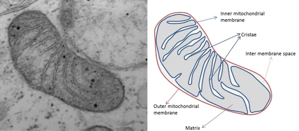
Here’s a electron microscope photo of a single mitochondrion (from this source).

What brain cells and muscle cells have in common is the need to maintain very different concentrations of potassium between themselves and the surrounding fluid (called a “gradient”), and that takes lots of energy.  So, any defect in mitochondria will tend to hurt brain cells and muscle cells first and worst.  In fact, childhood neurological dysfunction and muscle weakness are the two main features of a whole category of diseases caused by single-gene mutations affecting proteins used only by mitochondria.

In PSP, the mitochondrial problem is more subtle, but we don’t know exactly what it is or what causes it.  Here are some strands of evidence:

  • Brain cells growing in a dish that have had their own mitochondria destroyed and replaced by mitochondria isolated from blood cells of people with PSP don’t recover from various kinds of stress as well the same brain cells with replacement mitochondria from healthy people. 
  • Toxins damaging an important series of chemical reactions in the mitochondria called Complex I can cause a PSP-like condition in lab animals. 
  • Complex I and other components of mitochondria are also damaged by tau molecules with an abnormal number or location of attached phosphate molecules (“phospho-tau”), which we know occur in PSP.  The net effect is excessive levels of “free radicals,” which are toxic by-products of normal respiration.
  • While the most important gene mutation contributing to PSP risk is in MAPT, which encodes tau, the next-most important is PERK (protein kinase RNA-like endoplasmic reticulum kinase), which regulates the responses to stress in mitochondria.
  • Coenzyme Q-10, a nutritional supplement that assists Complex I, may help some of the immediate symptoms of PSP, as shown by at least one double-blind trial.

All the above is simply background justification to suspect that muscles and not just brain should be involved in PSP.  But there’s more direct evidence, too:

  • Muscle weakness and fatigue are more common in PSP than in others of the same age.
  • Weight loss is common in PSP and occurs early in the disease course.  The same is true for both in Parkinson’s, but not as markedly.
  • Grip strength is impaired in PSP.  That could be a result of changes in the brain, but the duration of the muscle fiber contractions is prolonged in PSP, a sign of muscle dysfunction.
  • Men (but, oddly, not women) with PSP have a reduced overall muscle mass relative to others of the same age. 
  • Muscle biopsy in people with PSP shows modest evidence of the same severe change in mitochondria (called “ragged red fibers”) that occur in the genetic mitochondrial diseases of childhood.

So, what’s the take-home for people with PSP? 

  • First, EXERCISE – including low-intensity muscle-building exercises.  Discuss the details first with your neurologist or physical therapist, and probably also with your primary care physician to make sure your heart and lungs are up to the task. 
  • Second, HAVE HOPE that insights into the mitochondrial role in PSP will bring new treatment or neuroprotection targeted at those cellular processes in the brain.  In fact, one such medication, called AMX-0035 (a combination of taurursodiol and sodium phenylbutyrate) will be entering a Phase 3 trial for PSP in the next few months.  The combination under the brand name “Relyvrio” was approved last year by the FDA for Lou Gehrig disease, where there’s a similar mitochondrial problem, so I have very high hopes that the same will happen for PSP.

Six horsemen of the Apocalypse

I watched a scientific presentation today in which the speaker started off by summarizing the leading theories of PSP’s pathogenesis.  That means not the external influences such as the genes received from one’s parents or whatever toxins or other stresses might help cause PSP in susceptible people.  Rather, it means the abnormal processes set in motion and operating inside in the brain cells leading to their dysfunction and eventually, their death. 

Here’s a quick rundown for you:

  • Tau splicing.  The tau protein is encoded by the MAPT gene, which has 14 sections called exons encoding separate fragments of the final protein.  These protein fragments are then stitched together, but sometimes one or more of them is omitted by design.  In healthy people, the product of exon 10 is included in about half of the final tau molecules, but in the tau tangles of PSP, that fragment is nearly always included.  This makes the tau more likely to aggregate.
  • Tau post-translational modifications. Many or most proteins have very small molecules attached to them at specific points to regulate their function and direct their folding pattern.  The abnormal tau of PSP has phosphate and other molecules in inappropriate places.  This could help explain the abnormal folding, which in turn produces toxic aggregates.
  • Tau degradation. The normal “garbage disposal” systems of brain cells gets rid of proteins or organelles (the tiny structures in cells that perform specific functions) that are either overproduced, defective or just worn out.  There are two basic kinds of such systems, the ubiquitin-proteasome system and the autophagy-lysosomal system.  Neither works as well as it should in PSP.  This allows abnormal tau and other toxic molecules to accumulate.
  • Intracellular tau spread. In many neurodegenerative diseases, the abnormally folded tau can travel from one brain cell to another, causing normal copies of those molecules to misfold in a similar fashion.  This creates a kind of chain reaction spreading the damage widely. The misfolding pattern of the tau is specific to each of the tauopathies.
  • Mitochondrial dysfunction. The mitochondria are the organelles in the cells that harvest energy from sugars with the help of oxygen.  In PSP, they function abnormally, possibly because of their own genetic mutations, possibly because their biochemistry is particularly sensitive to certain toxins in our environment.  Mitochondrial dysfunction doesn’t just deprive the cell of energy – it also produces toxic compounds such as free radicals that damage other cell components.
  • Gene expression errors. The most recently discovered pathomechanism has to do with abnormal regulation of access of the cell’s protein-making machinery to the DNA “blueprint.” That process is normally regulated by proteins collectively called “chromatin,” which coat and intertwine with the DNA in the nucleus.   One way the abnormality might work is that abnormal chromatin permits inappropriate access to certain genes that stimulate the immune system, producing a harmful inflammatory reaction in the brain.

All of these pathogenetic mechanisms except the first are currently being addressed by drugs in advanced stages of the development pipeline.  I really don’t know which horse to put my money on.