Strains and veins

I’ve been thinking about PSP subtypes a lot lately, mostly because of last week’s report of an eleventh subtype, PSP-PF, comprising elements of the PSP-PI and PSP-F types.  See my recent post for more explanation.  I’ve read what I can about what causes the various subtypes to prefer slightly different parts of the brain.  The general thought on that right now is “tau strains.” 

Think of tau as a species, like the dog, and its strains as breeds.  Let’s not get into the molecular nature of the inter-strain differences or what produces them.  Instead, let’s recognize that those strains could theoretically underlie the differences among the 11 PSP subtypes by introducing differences in predilections for different groups of brain cells sharing a location or function.  But this week, another possibility emerged as an explanation of the subtypes’ brain-area preferences: abnormal venous circulation.

The study in Parkinsonism and Related Disorders performed brain MRIs and routine clinical office exams on 95 people with PSP.  Of those, 64 had one of the three “cortical” subtypes (PSP-speech/language, PSP-corticobasal syndrome, and PSP-frontal).  The other 31 had one of the “subcortical” subtypes (PSP-Parkinsonism, PSP-progressive gait freezing, PSP-postural instability).  There were also 50 healthy participants as controls. 

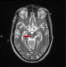
The three groups of participants were compared with regard to the size, number and location of any “white matter hyperintensities” (WMHs), examples of which appear in the MRIs below as the irregular white dots and splotches.  In mild form, they’re common in healthy, older people and more so in those with high blood pressure, diabetes and other vascular risk factors.  You can see how some of them sit smack up against the black slits in the middle of the brain, the spinal-fluid-filled lateral ventricles, and some are much closer to the outer, convoluted surfaces of the brain, the cerebral cortex.  (Image from Inzitari D, Pracucci G, Poggesi, et al. BMJ. 2009 Jul 6;339:b2477. doi: 10.1136/bmj.b2477

The graph below shows the current paper’s main results: (from Fu M-H, Satoh R, Ali F, et al. Parkinsonism and Related Disorders. 2025 Dec 22:143:108170. doi: 10.1016/j.parkreldis.2025.108170).

This graph’s vertical axis is a measure of the WMHs’ total volume, expressed as a percentage of total intracranial volume. The horizontal axis is the WMHs’ location expressed as average distance from the lateral ventricle.  The participants with the subcortical subtypes of PSP had the greatest volume of WMHs and their average distance from the lateral ventricles was greatest.  The people with the cortical subtypes ranked lower in both measures, and the control participants ranked lowest.  However, after correcting for various potential confounders, the differences remained statistically significant only for the areas between 12 and 30 mm from the lateral ventricles.

How to interpret this?  Let’s start with some background:

  • The tiny veins draining blood from the brain are divided into deep and superficial systems.  Each flows into its own set of larger veins en route to the heart. 
  • WMHs are areas of scarring. They’re largely of unknown cause, but they correlate with risk factors for stroke, which is mostly related to narrowing of arteries, not veins.
  • Multiple sclerosis, which produces areas of white matter inflammation and scarring more severe than those of PSP, has been linked to insufficiency of venous drainage of the brain.
  • Normal-pressure hydrocephalus, which is similar in many ways to PSP and even can have PSP-like changes in the brain cells, has been shown (by the same research group as the present paper) to include insufficiency in one of the deep veins.

The areas of brain yielding the graph’s statistically significant results drain into the deep venous system.  They’re unrelated to brain territories supplied by any specific arteries.  The authors tentatively conclude that the WMHs may be caused by insufficiency in the brain’s deep venous system.  They are appropriately cautious about assigning cause-and-effect, but the obvious question raised by their results is whether narrowing of the deep veins, and not any differences in tau or its post-translational modifications, could explain some, or maybe all, of the variety of PSP subtypes.

The authors of this paper overlap with those of last week’s about the new PSP-PF subtype summarized in my last post.  All from the Mayo Clinic in Rochester or Jacksonville, they include first author Dr. Mu-Hui Fu working under senior author Dr. Jennifer Whitwell, a veteran leader in PSP-related imaging research.

Good old MRI comes through

We still don’t have a great diagnostic test for PSP.  The best we can do is about 80%-90% sensitivity, specificity and positive predictive value.  In English:

  • Sensitivity is the fraction of people with PSP who give a positive result on the test.
  • Specificity is the fraction of people without PSP who give a negative result on the test.
  • Positive predictive value is the fraction of people with a positive test who actually have PSP.
  • A single number combining these into something useful in evaluating a single individual — rather than in comparing groups — is the “area under the receiver operating curve” (AUC; see this post for an explanation).  The AUC ranges from 0.50, which is no better than a coin toss, to 1.00, which is perfect accuracy.  An acceptable diagnostic test typically has an AUC of at least 0.85.

Most of the studies of PSP diagnostic markers have important weaknesses such as:

  • The studies frequently set up artificial situations such as distinguishing PSP only from PD or normal aging rather than from the long list of other possibilities that must be considered in the real world.  
  • The patients’ “true diagnoses” are usually defined by history and examination alone rather than by autopsy.
  • The patients included in the study were already known to have PSP by history and exam (or sometimes by autopsy), while the purpose of the marker would be to identify PSP in its much earlier, equivocal stages or in borderline or atypical cases.
  • The patients with PSP in most such studies are only those with PSP-Richardson’s syndrome, who account for only about half of all PSP in the real world.

The best type of marker so far is ordinary MRI.  Recently, a group of neurologists in Athens, Greece led by first author Dr. Vasilios C. Constantinides and senior author Dr. Leonidas Stefanis evaluated the specificity of various MRI-based measurements of brain atrophy.  One strength of their study was that their 441 subjects included people not only with PSP and Parkinson’s disease, but also with a long list of other conditions with which PSP is sometimes confused as well as a group of healthy age-matched controls. 

The single best MRI marker per this study was the area of the midbrain, the fat, V-shaped structure indicated below:

They found that MRI markers provided:

  • High diagnostic value (AUC >0.950 and/or sensitivity and specificity ∼90 %) to distinguish PSP from multiple system atrophy, Parkinson’s disease, and control groups.
  • Intermediate diagnostic value (AUC 0.900 to 0.950 and/or sensitivity and specificity 80 % to 90 %) to distinguish PSP from Alzheimer’s disease, frontotemporal dementia, dementia with Lewy bodies, and mild cognitive impairment (an early stage usually of AD).  
  • Insufficient diagnostic value (AUC < 0.900 or sensitivity/specificity ∼80 %) to distinguish PSP from corticobasal degeneration, normal-pressure hydrocephalus, and primary progressive aphasia (a language abnormality that can be caused by multiple specific diseases).
  • Insufficient value to distinguish the non-Richardson PSP subtypes from corticobasal degeneration and primary progressive aphasia, but good performance in the other comparators.

The researchers also concluded that:

  • One MRI measurement isn’t best for all the possible PSP comparators. 
  • Sometimes a combination of two or three measurements performed better than any single measurement.

One weakness of their method was the use of subjects diagnosed by standard history/exam (i.e., “clinical”) criteria, rather than by autopsy. Another is that their patients with PSP had had symptoms for an average of three years, so these were not subtle or early-stage cases. A letter to the journal’s editor from Dr. Bing Chen of Qingdao City, China further pointed out that the study of Constantinides and colleagues failed to account for the subtle effects of neurological medications on brain atrophy.  As PSP and the comparator disorders may be treated with different sets of drugs, taking this factor into account might enhance or reduce the apparent diagnostic value of MRI atrophy measurements.  

So, bottom line?  Drs. Constantinides and colleagues have given us the first study of MRI markers in PSP to include meaningful numbers of subjects with non-Richardson subtypes.  It’s also one of the few studies of any kind of PSP marker to include comparison of PSP a wide range of diagnostic “competitors” beyond just Parkinson’s and healthy aged persons.  Another plus is that the test, routine MRI, is nearly universally available, relatively inexpensive, and non-invasive.

The hope is that Pharma companies or others with candidate drugs will now have fewer or lower hurdles in the way of initiating clinical trials.

Orphans, this could be your chance

Back in 2023, I posted an explanation of the ten PSP subtypes.  The archetypal subtype, PSP-Richardson syndrome accounts for about half of all PSP and, in contrast to most of the other subtypes, has a rapid progression rate, a validated rating scale, and highly accurate diagnostic criteria.  All of these features have led clinical trial sponsors to maximize their trials’ sensitivity and minimize their costs by restricting admission to people with PSP-Richardson.  But developing better outcome measures for non-Richardson forms of PSP could change that practice.

A big step toward realizing this goal was published last week in the journal Neurology by a group at the Mayo Clinic in Rochester, MN.  Led by first author Dr. Mahesh Kumar and senior authors Drs. Jennifer Whitwell and Keith Josephs, the study found that a good outcome measure for clinical neuroprotection trials in all PSP subtypes was to combine a measure of atrophy by MRI with a measure of clinical disability.  This is a major advance.

The researchers performed brain MRIs at the start and end of a one-year period in 88 people with PSP and 32 age-matched controls.  Of those with PSP, 50 had PSP-Richardson, 18 had “PSP-cortical” (three of the other nine subtypes) and 20 had “PSP-subcortical” (the other 6 of the subtypes).  They had to lump the non-Richardson subjects using their subtypes’ general anatomical predilections because most of the subtypes were too rare to analyze on their own.

Calculating how much each of ten important PSP-involved brain regions had atrophied over the one-year interval allowed the researchers to identify which region(s) might best serve as markers of progression for each of the three groups when coupled with standard clinical measures.  Those measures include such familiar instruments as the PSP Rating Scale and the Unified Parkinson’s Disability Rating Scale’s motor section as well as less familiar scales specific for cognition, gait, eye movement and speech.  All the scales were administered concurrently with each of the two MRIs. 

They expressed the sensitivity to one-year progression not by some abstract statistic, but by the number of patients needed in a double-blind trial to demonstrate with at least 80% certainty that patients on active drug enjoyed a 20% slowing of progression relative to the placebo group. (These specifications are typical for PSP clinical trials.)  The better the measure’s performance, the fewer patients are needed.

And the award for Best Performance by an Outcome Measure in a PSP Neuroprotection Trial goes to . . . a combination of the rate of atrophy of whatever brain region shrinks fastest in the patient’s specific subtype and the PSP Rating Scale score.

The real significance of this study’s result is that using an outcome measure customized to each participant’s PSP subtype could allow trials to enroll not just people with PSP-Richardson, but also those with any of the other subtypes.  That’s because the trial’s measure of success could be to compare each patient’s rate of progression during the trial to that of patients in the placebo group with the same PSP subtype. 

This could double the number of people eligible to enroll in PSP trials, which means cutting the enrollment period in half, with commensurate reduction in costs for the sponsor.  The hybrid measure is more sensitive to progression than the PSP Rating Scale alone, thereby reducing the number of patients required even more. 

Both factors could lower the financial barrier confronting a company hoping to mount a trial for a promising PSP drug.  That may be the most important bottleneck right now in the development of a treatment to prevent or slow the progression of PSP.s

That’s why this news is huge for PSP in general and for the “orphans” in particular.